SHORT COMMUNICATION

Rapid Detection of Escherichia coli in Fresh Foods Using a Combination of Enrichment and PCR Analysis

Yukyung Choi1,2, Sujung Lee1,2, Heeyoung Lee1,2, Soomin Lee1,2, Sejeong Kim1,2, Jeeyeon Lee1,2, Jimyeong Ha1,2, Hyemin Oh1,2, Yewon Lee1,2, Yujin Kim1,2, Yohan Yoon1,2,*
Author Information & Copyright
1Department of Food and Nutrition, Sookmyung Women’s University, Seoul 04310, Korea
2Risk Analysis Research Center, Sookmyung Women’s University, Seoul 04310, Korea
*Corresponding Author : Yohan Yoon Department of Food and Nutrition, Sookmyung Women’s University, Seoul 04310, Korea Tel: +82-2-2077-7585 Fax: +82-2-710-9479, E-mail: yyoon@sookmyung.ac.kr

© Copyright 2018 Korean Society for Food Science of Animal Resources. This is an Open-Access article distributed under the terms of the Creative Commons Attribution Non-Commercial License (http://creativecommons.org/licenses/by-nc/3.0/) which permits unrestricted non-commercial use, distribution, and reproduction in any medium, provided the original work is properly cited.

Received: May 01, 2018 ; Revised: Jul 15, 2018 ; Accepted: Jul 22, 2018

Published Online: Aug 31, 2018

Abstract

The objective of this study was to determine the minimum enrichment time for different types of food matrix (pork, beef, and fresh-cut lettuce) in an effort to improve Escherichia coli detection efficiency. Fresh pork (20 g), beef (20 g), and fresh-cut lettuce (20 g) were inoculated at 1, 2, and 3 Log CFU/g of Escherichia coli. Samples were enriched in filter bags for 3 or 5 h at 44.5°C, depending on sample type. E. coli cell counts in the samples were enriched in E. coli (EC) broth at 3 or 5 h. One milliliter of the enriched culture medium was used for DNA extraction, and PCR assays were performed using primers specific for uidA gene. To detect E. coli (uidA) in the samples, a 3–4 Log CFU/mL cell concentration was required. However, E. coli was detected at 1 Log CFU/g in fresh pork, beef, and fresh-cut lettuce after 5, 5, and 3-h enrichment, respectively. In conclusion, 5-h enrichment for fresh meats and 3-h enrichment for fresh-cut lettuce in EC broth at 44.5°C, and PCR analysis using uidA gene-specific primers were appropriate to detect E. coli rapidly in food samples.

Keywords: fresh meat; fresh-cut lettuce; Escherichia coli; enrichment; PCR

Introduction

Food hygiene and safety are a major concern in the food industry, and microbiological safety is a particular problem. Escherichia coli can act as an indicator for the presence of other pathogenic bacteria, and it is detected easily in foods such as pork, beef, and chicken. Thus, E. coli detection in foods is one of the most useful hygienic criteria (Scheinberg et al., 2017; Seo et al., 2010; Simancas et al., 2016). However, at present the conventional method for E. coli detection requires several days (Feng et al., 2002; Stromberg et al., 2015; Wang and Salazar, 2016), especially in cases where E. coli concentrations are low. Enrichment is a commonly used method for bacterial isolation to increase the cell counts of target bacteria above other background flora prior to identification (Gracias and McKillip, 2004). According to FDA-BAM (U.S. Food and Drug Administration-bacteriological analytical manual) and other reports, E. coli can be enriched with E. coli (EC) broth or modified tryptic soy broth (mTSB); however, the enrichment methods are time-consuming (Feng et al., 2002; Stromberg et al., 2015).

Polymerase chain reaction (PCR), using primers against the uidA gene that encodes beta-D-glucuronidase can be used to identify E. coli accurately (Molina et al., 2015). PCR detection method has been used to identify a colony on an agar plate, which was formed by plating at least 24-h enriched broth. However, applying PCR detection method directly to the enriched samples has not been evaluated yet. In addition, there is an issue of specificity, since uidA gene is also present in Shigella (Frampton and Restaino, 1993). The objective of the present study was therefore to develop a rapid detection method for E. coli in food samples, using a combination of enrichment and PCR that can also differentiate E. coli from Shigella.

Materials and Methods

Bacterial preparation and determination of detection limit

Five E. coli strains (E. coli NCCP11142, E. coli NCCP14037, E. coli NCCP14038, E. coli NCCP14039, and E. coli NCCP15661), and Shigella sonnei NCCP14743 strain were cultured in 10 mL tryptic soy broth (TSB, Becton, Dickinson and Company, USA). One-hundred microliter aliquots were transferred to fresh 10 mL TSB, followed by incubation at 37°C for 24 h. The cultures of the five E. coli strains were mixed. Twenty-five milliliters of the E. coli mixture and 10 mL S. sonnei were centrifuged at 1,912 g and 4°C for 15 min, and the pellets were washed twice with the same volume of phosphate-buffered saline (PBS; 0.2 g KH2PO4, 1.5 g Na2HPO4, 8.0 g NaCl, and 0.2 g KCl in 1 L distilled H2O [pH 7.4]). The suspension was diluted with PBS to obtain 3, 4, and 5 Log CFU/mL of inocula, and E. coli and S. sonnei were assayed by PCR to determine the detection limit.

Food sample preparation and inoculation

Ham of pork and round of beef were purchased from a butcher shop, and a fresh-cut lettuce was purchased from a supermarket, located in Seoul, South Korea. Ham of pork and round of beef were cut into 20-g portions with a flame-sterilized knife. Fresh pork (20 g, n=4), beef (20 g, n=4), and fresh-cut lettuce (20 g, n=4) were placed aseptically into separate filter bags (3M, St. Paul, MN, USA). E. coli inoculum (0.1 mL) was inoculated onto the surface of the food samples to achieve 1, 2 and 3 Log CFU/g, and samples were massaged 20 times by hand. Samples were then left at room temperature (25°C) for 15 min to allow cell attachment.

E. coli enrichment in food samples

Eighty milliliters of EC broth (BD, USA) were placed into the filter bags, and shaken by hand 30 times. All samples were incubated at 44.5°C (Feng et al., 2002) for 0, 4, and 5 h for pork and beef, or 0 and 3, 6, 12 h for fresh-cut lettuce. After enrichment, 1-mL aliquots of the enriched samples were plated onto E. coli/coliform petrifilm (3M, USA) to quantify E. coli. The plates were incubated at 37°C for 24 h, and colonies were manually counted.

DNA extraction

One-milliliter aliquots of inocula and enriched samples were centrifuged at 18,341×g at 4°C for 5 min, and supernatants were discarded. Cell pellets were resuspended in 30 µL distilled water and boiled at 100°C for 10 min, and the suspensions were centrifuged at 18,341×g and 4°C for 3 min. The supernatants were then used for PCR analysis.

PCR analysis

Primers targeting the uidA and Shigella identification gene were used to differentiate E. coli from Shigella (Table 1). PCR conditions were as follows: 94°C for 2 min (initial denaturation), 94°C for 20 s (denaturation), 72°C for 20 s (extension), and 72°C for 2 min (final extension). Annealing was performed at 53°C for uidA or at 62°C for the Shigella identification gene for 10 s, and 35 cycles were performed. PCR analysis was performed using Fast mix French PCR (i-Taq) (iNtRon Biotechnology, Gyeonggi-do, Korea), and PCR products were run on an agarose gel (1.5%) with electrophoresis for 20 min. Target bands were visualized under UV light.

Table 1. Primers for polymerase chain reaction (PCR) analysis
Bacteria Target gene Size (bp) Primer sequence (5′-3′) Reference
Escherichia coli uidA 252 PT-2, GCGAAAACTGTGGAATTGGG
PT-3, TGATGCTCCATAACTTCCTG
Cebula et al. (1995)
Shigella Shigella identification gene 159 F255, TCGCATTTCTCTCCCCACCACG
F413, CCGGATGTGTCTCGGGCAATC
Kim et al. (2017)
Download Excel Table

Results and Discussion

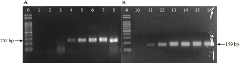
Minimum cell counts for PCR analysis, using primers for uidA gene were 3–4 Log CFU/mL for E. coli and 3 Log CFU/mL for Shigella (Fig. 1). From this result, we confirmed that 3–4 Log CFU/mL of bacterial cell counts was required to detect E. coli with primers targeting uidA gene, and the primers can detect both E. coli and Shigella. Hence, additional primers were necessary to differentiate E. coli from Shigella. Subsequently, the Shigella identification primers described in Table 1 were used, and the Shigella identification primers differentiated E. coli from Shigella (Fig. 1).

kosfa-38-4-829-g1
Fig. 1. Detection of Escherichia coli (A, uidA without enrichment; lanes 1–8) and Shigella (B, Shigella identification gene without enrichment; lanes 10–16) by PCR. Lanes 0 and 9: 100-bp ladder; lane 1: 1 Log CFU/mL cell counts; lanes 2 and 10: 2 Log CFU/mL cell counts; lanes 3 and 11: 3 Log CFU/mL cell counts; lanes 4 and 12: 4 Log CFU/mL cell counts; lanes 5 and 13: 5 Log CFU/mL cell counts; lanes 6 and 14: 6 Log CFU/mL cell counts; lanes 7 and 15: 7 Log CFU/mL cell counts; lanes 8 and 16: 8 Log CFU/mL cell counts.
Download Original Figure

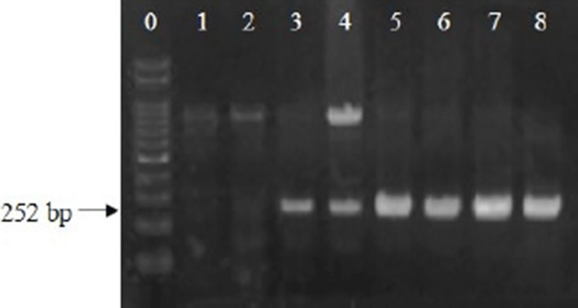
Analysis was then performed to determine optimum enrichment times required to obtain 3–4 Log CFU/mL of E. coli for PCR analysis. E. coli was inoculated into fresh pork, beef, or fresh-cut lettuce at 1, 2, and 3 Log CFU/g. E. coli in the pork and beef were enriched for 4 and 5 h, and E. coli in the fresh-cut lettuce were enriched for 3, 6 and 12 h. After 5-h enrichment, E. coli cell counts in the pork and beef increased to 5.9–6.0, 7.1, and 8.0–8.5 Log CFU/g for 1, 2, and 3-Log CFU/g inoculation levels, respectively, and uidA gene expression could be detected at all cell concentrations (Table 2, Fig. 2). In fresh-cut lettuce after 3-h enrichment, the bacterial cell counts increased to 4.2, 5.5, and 6.5 Log CFU/g for 1, 2, and 3-Log CFU/g inoculation levels, respectively, and uidA gene was positive for all samples (Table 2, Fig. 3). Thus, the optimal enrichment time for PCR detection of E. coli was 5 h for fresh pork and beef, and 3 h for fresh-cut lettuce.

Table 2. Escherichia coli cell counts (Log CFU/g, mean±SD) in fresh meats (pork and beef) and fresh-cut lettuce after 0, 4, and 5 h- and 0, 3, 6, and 12 h-enrichment with E. coli (EC) broth
Food matrix Targeted
E. coli cell counts
(Log CFU/g)
Enrichment time (h)
0 3 4 5 6 12
Pork 1 0.7±0.0 - 4.7±0.0 5.9±0.5 - -
2 1.5±0.0 - 5.8±0.0 7.1±0.4 - -
3 3.1±0.0 - 6.9±0.0 8.5±0.1 - -
Beef 1 0.8±0.2 - 4.4±0.6 6.0±0.6 - -
2 1.5±0.2 - 6.1±0.1 7.1±0.6 - -
3 3.0±0.0 - 7.3±0.1 8.0±0.2 - -
Fresh-cut lettuce 1 1.0±0.2 4.2±0.2 - - 6.3±0.0 7.2±0.0
2 1.7±0.2 5.5±0.2 - 8.3±0.0 9.0±0.0
3 3.1±0.2 6.5±0.1 - - 8.4±0.0 8.0±0.0

1) Not applied.

Download Excel Table
kosfa-38-4-829-g2
Fig. 2. Detection of Escherichia coli in fresh pork (A) and beef (B) samples by PCR for uidA after 5-h enrichment with E. coli (EC) broth. Lanes 0 and 9: 100-bp ladder; lanes 1, 2, 10, and 11: non-inoculated samples; lanes 3, 4, 12, and 13: 1-Log CFU/g inoculated samples; lanes 5, 6, 14, and 15: 2-Log CFU/g inoculated samples; lanes 7, 8, 16, and 17: 3-Log CFU/g inoculated samples.
Download Original Figure
kosfa-38-4-829-g3
Fig. 3. Detection of Escherichia coli in fresh-cut lettuce samples by PCR for uidA after 3-h enrichment with E. coli (EC) broth. Lane 0: 100-bp ladder; lanes 1 and 2: non-inoculated samples; lanes 3 and 4: 1-Log CFU/g inoculated samples; lanes 5 and 6: 2-Log CFU/g inoculated samples; lanes 7 and 8: 3-Log CFU/g inoculated samples.
Download Original Figure

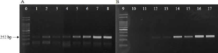
The above results show that meat samples require a longer enrichment time than fresh-cut lettuce. Low E. coli concentrations (0.7–0.8 Log CFU/g) in pork and beef increased to 4.4–4.7 Log CFU/g after 4-h enrichment, and the samples were negative for uidA expression (Table 2). However, at similar E. coli concentrations in fresh-cut lettuce, the samples were uidA positive. It is possible that a component of the meat samples is interfering with the PCR analysis. Wang and Salazar (2016) showed that a number of intrinsic factors can interfere with PCR assays, and other studies have shown that particulates such as fats and carbohydrates can affect nucleic acid amplification (Dwivedi and Jaykus, 2011; Thomas et al., 1991). For this reason, extra pre-treatment, such as centrifugation and bead-based techniques, are necessary to remove some particles from certain foods (Rossen et al., 1992; Yang et al., 2007). Heidenreich et al. (2010) detected E. coli in ground beef using an electrochemical biochip method after enrichment for 4–5 h, and Li et al. (2017) used propidium monoazide treatment to detect viable cell counts of E. coli O157:H7 at 12-h enrichment. However, in this present study, 5-h enrichment for fresh meat samples and 3-h enrichment for fresh-cut lettuce were sufficient to detect E. coli by PCR.

In conclusion, the combination of enrichment and PCR detection method is able to detect E. coli via applying PCR with uidA primers to samples directly after 5-h enrichment for fresh meats (pork and beef) and 3-h enrichment for fresh-cut lettuce.

Acknowledgements

This work was supported by the “Cooperative Research Program for Agriculture Science & Technology Development (Project No. PJ01193002)” Rural Development Administration, Korea.

References

1.

Cebula TA, Payne WL, Feng P. Simultaneous identification of strains of Escherichia coli serotype O157:H7 and their Shiga-like toxin type by mismatch amplification mutation assay-multiplex PCR. J Clin Microbiol. 1995; 33:248-250.

2.

Dwivedi HP, Jaykus LA. Detection of pathogens in foods: The current state-of-the-art and future directions. Crit Rev Microbiol. 2011; 37:40-63.

3.

Feng P, Stephen D, Weagant SD, Grant GA, Burkhardt W. Food Drug Administration (FDA)-Bacteriological Analytical Manual (BAM) chapter 4: Enumeration of Escherichia coli and the coliform bacteria. 2002Available from: https://www.fda.gov/Food/FoodScienceResearch/LaboratoryMethods/ucm064948.htm. Accessed at Mar 4, 2018

4.

Frampton EW, Restaino L. Methods for Escherichia coli identification in food, water and clinical samples based on beta-glucuronidase detection. J Appl Microbiol. 1993; 74:223-233.

5.

Gracias KS, McKillip JL. A review of conventional detection and enumeration methods for pathogenic bacteria in food. Can J Microbiol. 2004; 50:883-890.

6.

Heidenreich B, Poehlmann C, Sprinzl M, Gareis M. Detection of Escherichia coli in meat with an electrochemical biochip. J Food Prot. 2010; 73:2025-2033.

7.

Kim HJ, Ryu JO, Song JY, Kim HY. Multiplex polymerase chain reaction for identification of Shigellae and four Shigella species using novel genetic markers screened by comparative genomics. Foodborne Pathog Dis. 2017; 14:400-406.

8.

Li F, Li B, Dang H, Kang Q, Yang L, Wang Y, Aguilar ZP, Lai W, Xu H. Viable pathogens detection in fresh vegetables by quadruplex PCR. LWT Food Sci Technol. 2017; 81:306-313.

9.

Molina F, López-Acedo E, Tabla R, Roa I, Gómez A, Rebollo JE. Improved detection of Escherichia coli and coliform bacteria by multiplex PCR. BMC Biotechnol. 2015; 15:48.

10.

Rossen L, Nørskov P, Holmstrøm K, Rasmussen OF. Inhibition of PCR by components of food samples, microbial diagnostic assays and DNA-extraction solutions. Int J Food Microbiol. 1992; 17:37-45.

11.

Scheinberg JA, Dudley EG, Campbell J, Roberts B, DiMarzio M, DebRoy C, Cutter CN. Prevalence and phylogenetic characterization of Escherichia coli and hygiene indicator bacteria isolated from leafy green produce, beef, and pork obtained from farmers’ markets in Pennsylvania. J Food Prot. 2017; 80:237-244.

12.

Seo YH, Jang JH, Moon KD. Microbial evaluation of minimally processed vegetables and sprouts produced in Seoul, Korea. Food Sci Biotechnol. 2010; 19:1283-1288.

13.

Simancas A, Molina F, Tabla R, Roa I, Rebollo JE. YaiO, a new target for highly specific detection of Escherichia coli by PCR amplification. In Microbes in the spotlight: Recent progress in the understanding of beneficial and harmful microorganisms. In: Méndez-Vilas A, editor.Brown Walker Press. Boca Raton, FI, USA: 2016; p. 234.

14.

Stromberg ZR, Lewis GL, Marx DB, Moxley RA. Comparison of enrichment broths for supporting growth of Shiga toxin-producing Escherichia coli. Curr Microbiol. 2015; 71:214-219.

15.

Thomas EJ, King RK, Burchak JACK, Gannon VP. Sensitive and specific detection of Listeria monocytogenes in milk and ground beef with the polymerase chain reaction. Appl Environ Microbiol. 1991; 57:2576-2580.

16.

Wang Y, Salazar JK. Culture-independent rapid detection methods for bacterial pathogens and toxins in food matrices. Compr Rev Food Sci Food Saf. 2016; 15:183-205.

17.

Yang H, Qu L, Wimbrow AN, Jiang X, Sun Y. Rapid detection of Listeria monocytogenes by nanoparticle-based immunomagnetic separation and real-time PCR. Int J Food Microbiol. 2007; 118:132-138.