ARTICLE

Citrus sunki Peel Extract Enhances Proliferation and Differentiation of Fibro-Adipocyte Progenitors in Holstein Cattle for Cultivated Meat Production

Sehyuk Oh1,https://orcid.org/0000-0003-4105-2512, Gyutae Park1,https://orcid.org/0000-0003-1614-1097, Sanghun Park1https://orcid.org/0000-0003-4804-0848, Yunhwan Park1https://orcid.org/0000-0002-2239-6697, Nayoung Choi1https://orcid.org/0000-0002-4782-1098, Youngho Lim1https://orcid.org/0000-0002-0238-4736, Soyoung Jang1https://orcid.org/0009-0001-1146-2695, Youngjin Kim1https://orcid.org/0009-0002-6243-3250, Hyunsu Choi1https://orcid.org/0000-0002-7516-2536, Sungkyun Oh1https://orcid.org/0009-0006-4770-9537, Jungseok Choi1,*https://orcid.org/0000-0001-8033-0410
Author Information & Copyright
1Department of Animal Science, Chungbuk National University, Cheongju 28644, Korea
*Corresponding author : Jungseok Choi, Department of Animal Science, Chungbuk National University, Cheongju 28644, Korea, Tel: +82-43-261-2551, Fax: +82-43-261-2773, E-mail: jchoi@chungbuk.ac.kr

These authors contributed equally to this work.

© Korean Society for Food Science of Animal Resources. This is an Open-Access article distributed under the terms of the Creative Commons Attribution Non-Commercial License (http://creativecommons.org/licenses/by-nc/3.0) which permits unrestricted non-commercial use, distribution, and reproduction in any medium, provided the original work is properly cited.

Received: Dec 13, 2024 ; Revised: Feb 28, 2025 ; Accepted: Mar 04, 2025

Published Online: Nov 01, 2025

Abstract

The importance of fat cell culture is increasing in the process of cultured meat production to improve meat quality. Among them, intramuscular fat, which greatly affects its quality, is mainly derived from fibro-adipogenic progenitor cells (FAPs). In this study, the effect of Citrus sunki peel extract (CPE) on the proliferation and adipose differentiation of FAPs isolated from muscle of Holstein cattle was investigated to enhance the proliferation and differentiation abilities of FAPs. FAPs were cultured in basal medium (C) or basal medium supplemented with 0.05% dimethyl-sulfoxide (CDMSO) or basal medium supplemented with 50, 100, 200, 300, and 400 μg/mL CPE (CPE50, CPE100, CPE200, CPE300, and CPE400). Live cell counts of CPE100 was significantly higher than in C and CDMSO. The results of MTS assay revealed significantly higher levels of CPE50 and CPE100 than in C and CDMSO. In reverse transcription quantitative polymerase chain reaction and Western blot experiments, the gene expression (CEBPA, CEBPB and PPARG) and protein expression (FASN, CEBPB, and PPARG) in FAPs cultured with CPE was significantly higher than or comparable to C. In conclusion, the addition of CPE (especially 100 μg/mL) enhanced the proliferation and differentiation of FAP of Holstein cattle for cultured meat development.

Keywords: Citrus sunki peel; cultivated meat; differentiation; fibro-adipogenic progenitors; proliferation

Introduction

Cultivated meat technologies are environmentally sustainable and contribute to animal welfare compared with traditional meat production (Post, 2012; Stephens et al., 2018). Compared with traditionally produced beef, sheep and pork, the production of cultivated meat production uses approximately 82%–96% less water use, 78%–96% less greenhouse gas emissions, 7%–45% less energy use and involves 99% less land use (Stephens et al., 2018). Meat contains intramuscular fat (IMF). The IMF plays an important role in food quality, such as sensory properties and health. In general, IMF content has a positive effect on the sensory properties of meat such as flavor, juiciness, and tenderness (Hocquette et al., 2010). Since fat is a flavor compound and affects transient release of flavor (Elmore et al., 2002), increasing the IMF improves flavor (Thompson, 2004). Many cells that differentiate into fat are being studied to obtain IMF for cultivated meat. It is possible to differentiate fat for cultivated meat from various cells, such as dedifferentiated adipocytes, embryonic stem cells, and induced pluripotent stem cells (Fish et al., 2020; Hill et al., 2019; Wei et al., 2013). Among them, intramuscular adipose tissue is mainly derived from a stromal stem cell population known as fibro-adipogenic progenitors (FAPs; Guan et al., 2017; Huang et al., 2012; Loomis et al., 2022). FAPs are differentiated from satellite cells via platelet-derived growth factor receptor A (PDGFRA) expression (Fitzgerald et al., 2023; Joe et al., 2010; Uezumi et al., 2010). FAPs contribute to muscle regeneration under physiological conditions, and to fibrogenesis and adipogenesis during pathology (Heredia et al., 2013).

Citrus fruits contain beneficial phytochemicals (Satari and Karimi, 2018). It contains phenylpropanoids, coumarins, carotenoids, and flavonoids, including polymethoxylated flavones such as nobiletin, tangeretin, naringin, and hesperidin (Kang et al., 2005). Natural extracts derived from citrus fruits are rich in flavonoids and exhibit anti-inflammatory, antioxidant, and anti-cancer activities (Galati et al., 1994; Ko et al., 2010; Lu et al., 2010). Citrus sunki has been used in oriental medicine since ancient times (Kang et al., 2005). Nobiletin and tangeretin, which are unique components of citrus fruits, are particularly abundant in Citrus sunki, and have excellent antioxidant, anti-inflammatory, and anti-obesity effects (Kang et al., 2012; Pang et al., 2023; Shin et al., 2011). These bioactive substances are contained in the peel rather than the fruit pulp and are more physiologically effective (Chung et al., 2000; Wu et al., 2013; Yoshigai et al., 2013). However, one-third of citrus fruit is processed, and the large amount of peel produced during juice processing is generally considered agro-industrial waste (Negro et al., 2016). Incineration and landfilling for the disposal of citrus by-products, which are produced in tons per day, can cause environmental and economic problems (Casquete et al., 2015; Satari and Karimi, 2018). Increasing the utilization of citrus by-products may be a solution to the disposal problem. Few studies have investigated the role of natural products in enhancing the culture and differentiation of FAPs to IMF. Furthermore, the effects of citrus fruits on the differentiation of FAPs into fat have not been reported. The purpose of this study was to investigate the effect of Citrus sunki peel extract (CPE) on proliferation and differentiation of FAPs from Holstein cattle, a popular breed of cattle, in cultivated meat production.

Materials and Methods

Cell isolation and sorting

The Holstein cattle used in the experiment was approved by the Institutional Animal Care and Use Committees (IACUC) of Chungbuk National University (CBNUA-2257-24-01). The cells were isolated using collagenase type II (600 units/mL DMEM) and centrifugation from Holstein cattle's buttock muscle. The obtained cells were suspended in freezing media (Gibco, Waltham, MA, USA) and stored in liquid nitrogen. For sorting of cells using fluorescence-activated cell sorting (FACS), cells were suspended in FACS buffer [0.1% bovine serum albumin (BSA; Roche, Basel, New Zealand) in PBS]. The cells were then stained with CD29 antibody (APC, 303008, BioLegend, San Diego, CA, USA), and CD56 antibody (PE-CyTM7, 335826, BD Bioscience, Franklin Lakes, NJ, USA). FAPs were sorted into the CD29+/CD56- population using the FACS Aria II Cell Sorter (BD Bioscience).

Extraction of Citrus sunki peel

The Citrus sunki peel was broken into small pieces for extraction, and 50 g was extracted at 70°C for 6 h with 1 L 60% ethanol. It was then filtered through a 0.4 μm filter and concentrated using a rotary vacuum evaporator. The concentrated CPE was lyophilized and stored.

High-performance liquid chromatography analysis

The high-performance liquid chromatography (HPLC) analysis was performed to analyze the nobiletin content of Citrus sunki extract. The sample to be analyzed was 2.4 mg CPE to which 12 mL of ethanol was added and separated at 30°C using a column (150 mm×4.6 mm, 5 μm Zorbax Eclipse XDB-C18; Agilent, Santa Clara, CA, USA). The mobile phase was methanol and distilled water (pH adjusted to 3 using glacial acetic acid). The gradient elution program was as follows: 25%–40% methanol from 0–24 min, 40%–62% methanol from 24–35 min, 62% methanol from 35–44 min, 62%–80% methanol from 44–50 min, and 85%–100% methanol from 50–60 min. The detection wavelength was 330 nm.

Culture of fibro-adipogenic progenitors from Holstein cattle

FAPs for differentiation were cultured in Matrigel-coated flasks. Cells were seeded at 2,000 cells/cm2 and cultured in Ham's F-10 medium (20% FBS, 1% PSA) for 6 days in an incubator (37°C, 5% CO2). After more than 80% confluence, cells were differentiated in DMEM (11995-065, Gibco) containing 1% FBS, 1% PSA, 0.5 mM 3-isobutyl-1-methylxanthine (IBMX; I5879, Sigma-Aldrich, Burlington, MA, USA), 1 μM dexamethasone (D-085, Sigma-Aldrich), and 10 μM insulin (I0516, Sigma-Aldrich) for 3 days. Then, replace the DMEM medium containing 1% FBS, 1% PSA, and 10 μM insulin once every 3 days for a total of 6 days.

Cell counting and MTS assay

Cell counting was measured with a cell counter (Countess®, Invitrogen, Waltham, MA, USA) using trypan blue. The MTS assay was performed to measure cell number via mitochondrial activity. The MTS assay was measured using MTS solution (G3582, Promega, Madison, WI, USA), and absorbance was measured a wavelength at 490 nm following 2 h of incubation at 37°C.

Immunofluorescence staining and Oil Red O staining

Immunofluorescence staining of FAPs was fixed using 2% paraformaldehyde and permeabilized using 0.1% Triton-X (in PBS). Permeabilized cells were blocked using 2% BSA (Roche) and stained with PDGARA primary antibody and secondary antibody (anti-rabbit IgG cross-labeled antibody; Invitrogen). After antibody staining, nuclei were counterstained using Hoechst 33342 (H3570, Invitrogen) and images were measured. The differentiated FAPs were fixed by incubation with 2% paraformaldehyde for 40 min. Fixed cells were incubated in 60% isopropanol for 5 min and then stained using Oil Red O solution. The Oil Red O solution was prepared using a 2:3 ratio of distilled water:Oil Red O.

Reverse transcription quantitative polymerase chain reaction

RNA extraction was performed on day 6 of differentiation using TRIzol reagent, and Reverse Transcription Master Premix (Elpis-Biotech, Seongnam, Korea) was used to convert the extracted RNA into cDNA. The cDNA conversion was incubated at 60°C for 1 h and 94°C for 5 min. Reverse transcription quantitative polymerase chain reaction (RT-qPCR) was performed with 1 μL of primers and 1 μL of cDNA in a total volume of 20 μL using SYBR Green solution (A46109, Applied BiosystemsTM, Waltham, MA, USA). Table 1 lists the primer sequences used in the RT-qPCR.

Table 1. Primer sequences used in RT-qPCR
Primer Direction Sequence (5’→3’)
GAPDH F GGGTCATCATCTCTGCACCT
R GGTCATAAGTCCCTCCACGA
CEBPA F CTGGAGCTGACCAGTGACAA
R GGGATGGACTGATTGTGCTT
CEBPB F TACTACGAGGCGGACTGCTT
R GTTGCTCCACCTTCTTCTGG
PPARG F ATTTGGAAACGGACGTCTTG
R TGAGGTCCTTGCAGACACTG
FASN F CCCAGAGCTGGACTACTTCG
R GAGCCGTCAAACAGGAAGAG

RT-qPCR, reverse transcription quantitative polymerase chain reaction.

Download Excel Table
Western blot

Proteins from FAPs were collected using RIPA lysis buffer. The concentration of the obtained protein was measured using the Bradford assay and equilibrated. Proteins were separated by TGX Precast Gels (Bio-Rad Laboratories, Hercules, CA, USA), and the gel was transferred to a PVDF membrane. Affinity Purified Goat Anti-Mouse IgG (H+L) HRP-conjugated antibody was used as the secondary antibody.

Statistical analysis

All experiments were performed at least three times. The results were analyzed using the statistical program SAS (9.4 for Windows, SAS Institute, Cary, NC, USA) to determine significance (p<0.05) using the Duncan multiple range test.

Results and Discussion

Preparation of Holstein cattle fibro-adipogenic progenitors

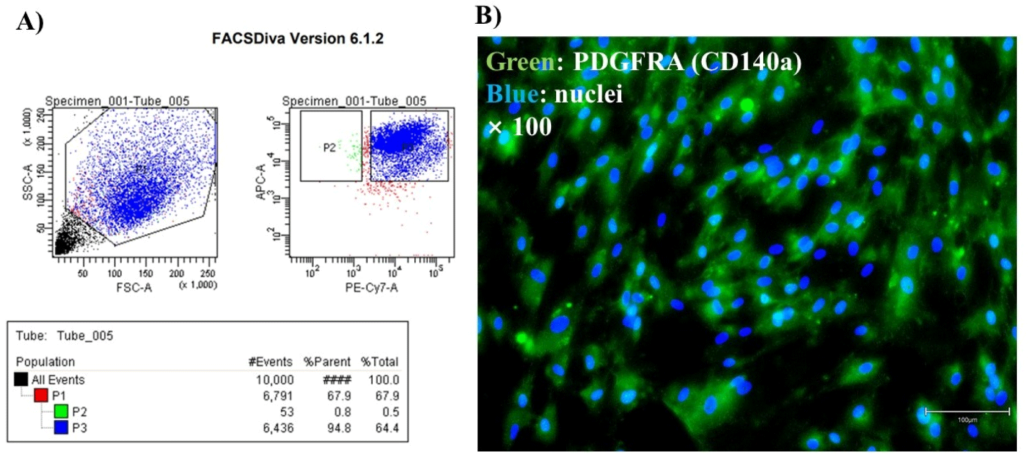
Cells from Holstein cattle muscle tissue were gated for the CD29+/56- population and these cells represented FAPs (Fig. 1A). To confirm that the cells sorted by FACS were FAPs, fluorescence staining was performed with a PDGFRA antibody (Fig. 1B). Progenitors of the non-myogenic lineage have the potential to become adipogenic (Joe et al., 2010; Wosczyna et al., 2012). PDGFRA is a specific surface marker for these non-myogenic precursor cells (also called FAPs; Uezumi et al., 2010). PDGFRA positivity has establishes the presence of FAPs in various experiments (Guan et al., 2017).
kosfa-45-6-1740-g1
Fig. 1. FACS and fluorescence staining results of FAPs in Holstein cattle. (A) Representative flow cytometry plots of unsorted bovine muscle cells, based on surface expression of CD29-APC and CD56-PE-Cy7. Cell in the P2 area represents FAPs (CD29+/CD56-). (B) Immunofluorescence staining was performed to identify FAPs using PDGFRA, a FAP marker, and Hoechst (×100). FACS, fluorescence-activated cell sorting; PDGFRA, platelet-derived growth factor receptor A; FAPs, fibro-adipogenic progenitors.
Download Original Figure
High-performance liquid chromatography analysis

Fig. 2A presents the CPE chromatogram and the nobiletin structure. The marked portion in Fig. 2B represents nobiletin (Li et al., 2021) and the area indicates 8,543.73 mVs. Substituting the standard curve, CPE contains 5,006.35 ppm of nobiletin.

kosfa-45-6-1740-g2
Fig. 2. Results of HPLC analysis of citrus peel extract. (A) Nobiletin concentration (ppm) relative to the area of HPLC chromatograms. (B) HPLC chromatograms of CPE. The marked part represents nobiletin and its chemical structure. HPLC, high-performance liquid chromatography; CPE, Citrus sunki peel extract.
Download Original Figure
Cell proliferation capacity

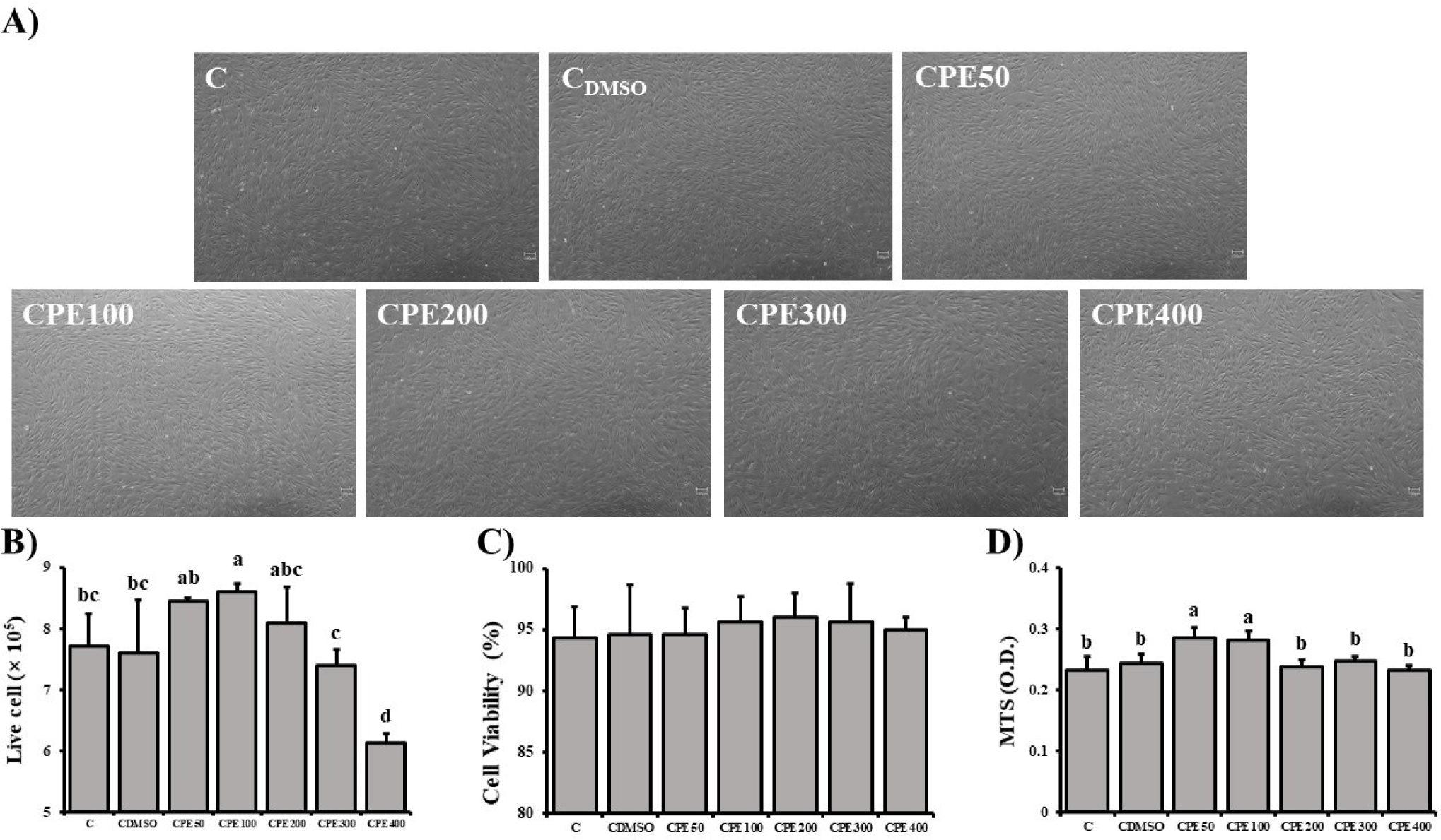
FAPs were cultured for 6 days in each experimental design (C, CDMSO, CPE50, CPE100, CPE200, CPE300, and CPE400). Since the concentration of DMSO in the medium with CPE was 0.05%, it was compared with the medium containing only 0.05% DMSO without CPE (Fig. 3A). The live cell count and viability of FAPs were measured (Figs. 3B and C). The live cell count of FAPs was higher in FAPs with 100 μg/mL CPE compared with control groups (p<0.05). CPE50 and CPE200 were significantly higher than CDMSO (p<0.05). Fig. 3D shows the results of the MTS analysis, where the FAP of CPE50 and 100 were significantly higher than the control group (p<0.05).
kosfa-45-6-1740-g3
Fig. 3. Experimental results (cell counting and MTT assay) demonstrating the proliferation capacity of Holstein cattle FAPs based on the amount of citrus peel extract added in the cultured medium. Values are mean±SD (n=3). a–d Different letters indicate statistically significant differences (p<0.05). (A) Image of control and treatment groups (×40; Scale bar=100 μm). (B) Live cells count of FAPs proliferation in 6 days. (C) Cell viability of FAPs proliferating for 6 days. (D) Results of MTS assay of FAP proliferation in 6 days. FAPs, fibro-adipogenic progenitors.
Download Original Figure

Reactive oxygen species (ROS) contribute to cell death, necrosis, and inhibition of cell proliferation (Matés et al., 2008). Excessive ROS induces cell death through activation of MAPK, AMPK, and BNI, inactivation of ATG4, and mitochondrial damage. Such ROS-induced cell death can be suppressed by antioxidant action (Villalpando-Rodriguez and Gibson, 2021). Flavonoids are antioxidants present in citrus peel and protect cells from free radical damage (Ashraf et al., 2017), and this effect is expected to increase the proliferative capacity of FAPs. Also, because citrus peel is rich in polyphenols, and its antioxidant capacity is higher than that of other edible fruits (Singh et al., 2020). In the study of Ikawati et al. (2019), C. reticulata extract at concentrations as low as 100 μg/mL induced cell proliferation, while higher concentrations decreased cell viability.

Differentiation capacity

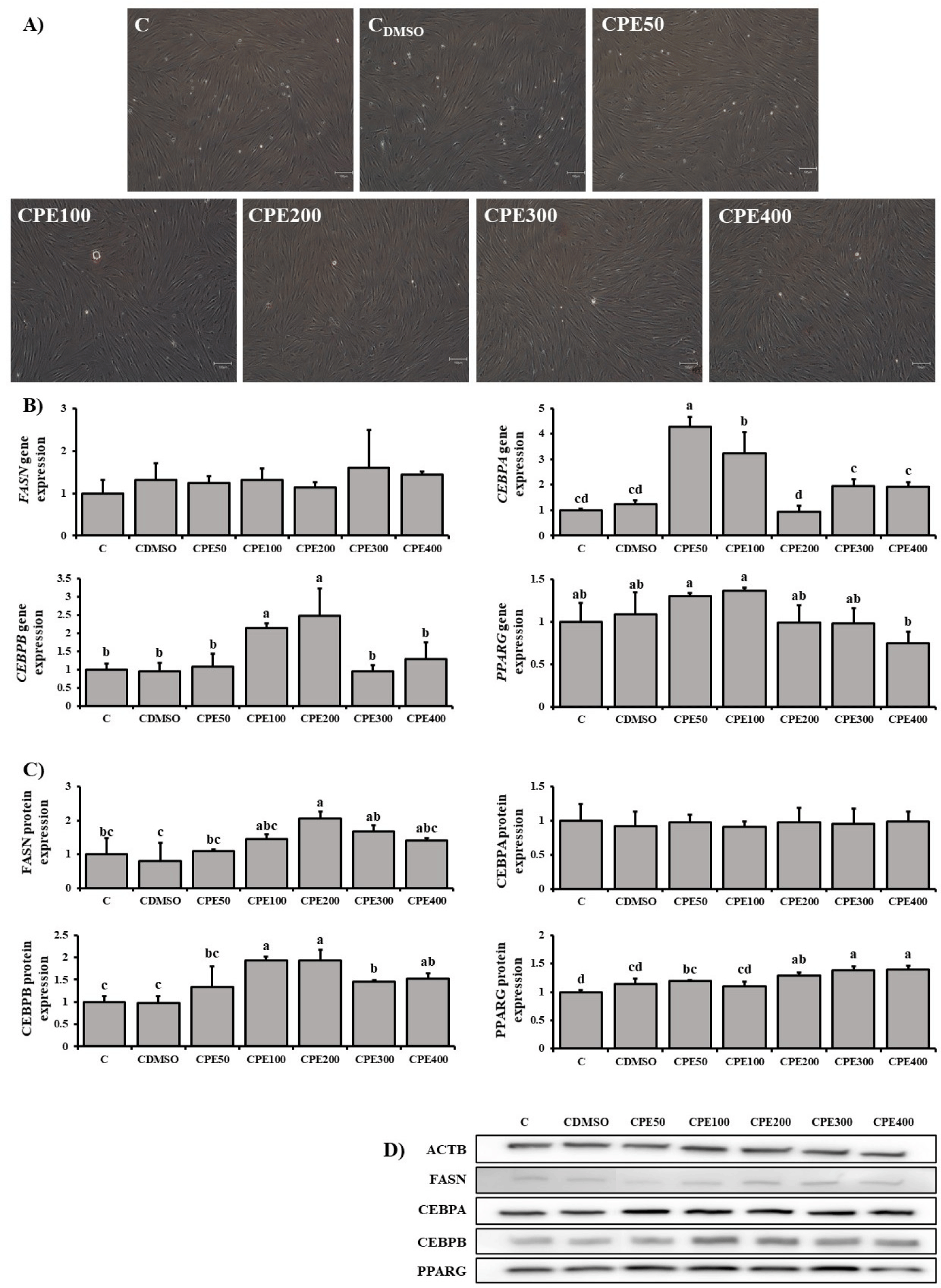
Differentiation of FAPs was confirmed by Oil Red O staining (Fig. 4A). RT-qPCR results of gene expression of FASN, CEBPA, CEBPB, and PPARG were compared (Fig. 4B). No significant difference in FASN gene expression was detected in all treatment and control groups. The expression of CEBPA gene was significantly higher in CPE50 and CPE100 compared with control groups (p<0.05). The expression of CEBPB gene was significantly higher in CPE100 and CPE200 than in control groups.
kosfa-45-6-1740-g4
Fig. 4. Experimental results (Oil Red O staining, RT-qPCR, and western blot) demonstrating the differentiation capacity of Holstein cattle FAPs according to the amount of citrus peel extract added to the cultured medium. Values are mean±SD (n=3). a–d Different letter indicate statistically significant differences (p<0.05). (A) Images of Holstein cattle FAPs that differentiated for 6 days with Oil Red O staining (×100; scale bar=100 μm). (B) The relative gene expression of FASN, CEBPA, CEBPB, and PPARG, adipogenesis-related gene, in Holstein cattle FAPs differentiating for 6 days in the medium containing different concentrations of Citrus sunki peel extract. (C) The relative expression of adipogenesis-related proteins (FASN, CEBPA, CEBPB, and PPARG) in Holstein cattle FAPs differentiating for 6 days in the medium containing different concentrations of CPE. (D) Western blot analysis of FASN, CEBPA, CEBPB, and PPARG expression. RT-qPCR, reverse transcription quantitative polymerase chain reaction; FAPs, fibro-adipogenic progenitors.
Download Original Figure

The protein expression of FASN, CEBPA, CEBPB, and PPARG was compared via Western blotting analysis (Fig. 4C).

The expression of FASN protein was significantly higher in CPE200 compared with control groups (p<0.05). The CEBPB protein expression in the other treatment groups varied significantly from that of the control groups (p<0.05), and the difference was the greatest in CPE100 and CPE200. The expression of PPARG protein was significantly higher in CPE300 and CPE400 than in control groups.

The CEBP family members exert a multifaceted influence on the differentiation of preadipocytes, playing a pivotal role in the processes of adipogenesis and differentiation. The transcription factors CEBPB and CEBPG are the initial mediators of differentiation in preadipocytes following exposure to differentiation factors, thereby initiating the differentiation process (Darlington et al., 1998). During the process of adipocyte differentiation, the transcription factors and CEBPD and CEBPB induce the expression of CEBPA (Rosen and MacDougald, 2006). As reported by Lin and Lane (1992), the inhibition of CEBPA expression in preadipocytes resulted in the absence of triacylglycerol accumulation and the lack of expression of fat-specific genes. This evidence supports the assertion that CEBPA is a crucial factor in the differentiation of preadipocytes. CEBPB induces CEBPA and acts as a transcriptional regulator of PPARG, a master regulator of adipogenesis (Hamm et al., 2001; Rosen et al., 2000). Adipogenesis is regulated not only by CEBP but also by PPARG (Gupta et al., 2010; Spiegelman and Flier, 1996). In addition, PPARG is required for adipogenesis and maintenance of differentiation status (Imai et al., 2004). PPARG is also important for adipocyte function, including insulin sensitivity, adipokine secretion, and lipid metabolism (Rangwala and Lazar, 2004). Consequently, two transcription factors in preadipocytes, CEBPA and PPARG, have been characterized as critical regulators of adipogenic differentiation (Lin and Lane, 1994; Tontonoz et al., 1994). The mutual expression of CEBPA and PPARG is enhanced, and these factors promote the differentiation of adipose tissue and increase the accumulation of lipids (Rosen et al., 2002; Tanaka et al., 1997). PPARG plays an important role in IMF in cattle, and the expression of CEBPA and PPARG in skeletal muscle is higher in cattle breeds with a high capacity for IMF deposition (Duarte et al., 2013; Moisá et al., 2014). FASN represents a delayed adipogenic marker and is a pivotal enzyme in the metabolic pathway of fatty acids. (Habinowski and Witters, 2001; Kim and Spiegelman, 1996).

The results of this study showed that CPE positively regulates CEBPA, CEBPB, PPARG, and FANS during the differentiation of FAPs. Another study showed that nobiletin increased the CEBPB expression of 3T3-L1 cells (Saito et al., 2007). The activation of extra cellular signal-regulated kinase and cAMP-responsive element-binding protein enhanced by nobiletin activated CEBPB and PPARG to promote adipogenesis (Saito et al., 2007). In addition to nobiletin, tangeretin makes up a large portion of citrus peel and is a potential insulin sensitizer (Guo et al., 2020). In addition to nobiletin, citrus fruits contain a variety of other flavonoids that enhance and positively affect adipogenesis (Bae et al., 2022; Kuroyanagi et al., 2008; Onda et al., 2013). Therefore, it is thought that CPE may enhance adipogenesis.

Conclusion

In our study, CPE enhanced the proliferation and differentiation of Holstein cattle FAPs and resulted in positive effects. In particular, the addition of 100 μg/mL of CPE was the most effective in terms of proliferation and differentiation induction for efficient cell culture-based meat production.

Conflicts of Interest

The authors declare no potential conflicts of interest.

Acknowledgements

This research was supported by Korea Institute of Planning and Evaluation for Technology in Food, Agriculture and Forestry (IPET) through the High Value-added Food Technology Development Project, funded by Ministry of Agriculture, Food and Rural Affairs (MAFRA) (321028-5). This work was supported by “Regional Innovation Strategy (RIS)” through the National Research Foundation of Korea (NRF) funded by the Ministry of Education (MOE) (2021RIS-001).

Author Contributions

Conceptualization: Oh S, Park G, Jang S. Data curation: Oh S, Park G, Park Y, Choi N, Lim Y, Jang S. Formal analysis: Oh S, Park G, Park S, Jang S, Choi H. Methodology: Oh S, Park G, Park S, Park Y, Choi N, Jang S, Choi H. Software: Oh S, Park G, Park Y, Lim Y, Kim Y, Choi H. Validation: Oh S, Park G, Lim Y, Jang S, Oh S, Choi J. Investigation: Oh S, Park G, Park S, Park Y, Choi H, Oh S. Writing - original draft: Oh S, Park G, Choi J. Writing - review & editing: Oh S, Park G, Park S, Park Y, Choi N, Lim Y, Jang S, Kim Y, Choi H, Oh S, Choi J.

Ethics Approval

All animal studies were performed in accordance with international regulations of the usage and welfare of laboratory animals and protocols approved by the Institutional Animal Care and Use Committee (IACUC) in Chungbuk National University in terms of ethic procedures and scientific care (CBNUA-2257-24-01).

References

1.

Ashraf H, Butt MS, Iqbal MJ, Suleria HAR. 2017; Citrus peel extract and powder attenuate hypercholesterolemia and hyperglycemia using rodent experimental modeling. Asian Pac J Trop Biomed. 7:870-880

2.

Bae J, Yang Y, Xu X, Flaherty J, Overby H, Hildreth K, Chen J, Wang S, Zhao L. 2022; Naringenin, a citrus flavanone, enhances browning and brown adipogenesis: Role of peroxisome proliferator-activated receptor gamma. Front Nutr. 9:1036655

3.

Casquete R, Castro SM, Martín A, Ruiz-Moyano S, Saraiva JA, Córdoba MG, Teixeira P. 2015; Evaluation of the effect of high pressure on total phenolic content, antioxidant and antimicrobial activity of citrus peels. Innov Food Sci Emerg Technol. 31:37-44

4.

Chung SK, Kim SH, Choi YH, Song EY, Kim SH. 2000; Status of citrus fruit production and view of utilization in cheju. Food Ind Nutr. 5:42-52.

5.

Darlington GJ, Ross SE, MacDougald OA. 1998; The role of C/EBP genes in adipocyte differentiation. J Biol Chem. 273:30057-30060

6.

Duarte MS, Paulino PVR, Das AK, Wei S, Serão NVL, Fu X, Harris SM, Dodson MV, Du M. 2013; Enhancement of adipogenesis and fibrogenesis in skeletal muscle of Wagyu compared with Angus cattle. J Anim Sci. 91:2938-2946

7.

Elmore JS, Campo MM, Enser M, Mottram DS. 2002; Effect of lipid composition on meat-like model systems containing cysteine, ribose, and polyunsaturated fatty acids. J Agric Food Chem. 50:1126-1132

8.

Fish KD, Rubio NR, Stout AJ, Yuen JSK, Kaplan DL. 2020; Prospects and challenges for cell-cultured fat as a novel food ingredient. Trends Food Sci Technol. 98:53-67

9.

Fitzgerald G, Turiel G, Gorski T, Soro-Arnaiz I, Zhang J, Casartelli NC, Masschelein E, Maffiuletti NA, Sutter R, Leunig M, Farup J, De Bock K. 2023; MME+ fibro-adipogenic progenitors are the dominant adipogenic population during fatty infiltration in human skeletal muscle. Commun Biol. 6:111

10.

Galati EM, Monforte MT, Kirjavainen S, Forestieri AM, Trovato A, Tripodo MM. 1994; Biological effects of hesperidin, a citrus flavonoid (note i): Antiinflammatory and analgesic activity. Farmaco (Soc Chim Ital 1989). 40:709-712.

11.

Guan L, Hu X, Liu L, Xing Y, Zhou Z, Liang X, Yang Q, Jin S, Bao J, Gao H, Du M, Li J, Zhang L. 2017; bta-miR-23a involves in adipogenesis of progenitor cells derived from fetal bovine skeletal muscle. Sci Rep. 7:43716

12.

Guo J, Chen J, Ren W, Zhu Y, Zhao Q, Zhang K, Su D, Qiu C, Zhang W, Li K. 2020; Citrus flavone tangeretin is a potential insulin sensitizer targeting hepatocytes through suppressing MEK-ERK1/2 pathway. Biochem Biophys Res Commun. 529:277-282

13.

Gupta RK, Arany Z, Seale P, Mepani RJ, Ye L, Conroe HM, Roby YA, Kulaga H, Reed RR, Spiegelman BM. 2010; Transcriptional control of preadipocyte determination by Zfp423. Nature. 464:619-623

14.

Habinowski SA, Witters LA. 2001; The effects of AICAR on adipocyte differentiation of 3T3-L1 cells. Biochem Biophys Res Commun. 286:852-856

15.

Hamm JK, Park BH, Farmer SR. 2001; A role for C/EBPβ in regulating peroxisome proliferator-activated receptor γ activity during adipogenesis in 3T3-L1 preadipocytes. J Biol Chem. 276:18464-18471

16.

Heredia JE, Mukundan L, Chen FM, Mueller AA, Deo RC, Locksley RM, Rando TA, Chawla A. 2013; Type 2 innate signals stimulate fibro/adipogenic progenitors to facilitate muscle regeneration. Cell. 153:376-388

17.

Hill ABT, Bressan FF, Murphy BD, Garcia JM. 2019; Applications of mesenchymal stem cell technology in bovine species. Stem Cell Res Ther. 10:44

18.

Hocquette JF, Gondret F, Baéza E, Médale F, Jurie C, Pethick DW. 2010; Intramuscular fat content in meat-producing animals: Development, genetic and nutritional control, and identification of putative markers. Animal. 4:303-319

19.

Huang Y, Das AK, Yang QY, Zhu MJ, Du M. 2012; Zfp423 promotes adipogenic differentiation of bovine stromal vascular cells. PLOS ONE. 7e47496

20.

Ikawati M, Armandari I, Khumaira A, Ertanto Y. 2019; Effects of peel extract from citrus reticulata and hesperidin, a citrus flavonoid, on macrophage cell line. Indones J Pharm. 30:260-268

21.

Imai T, Takakuwa R, Marchand S, Dentz E, Bornert JM, Messaddeq N, Wendling O, Mark M, Desvergne B, Wahli W, Chambon P, Metzger D. 2004; Peroxisome proliferator-activated receptor γ is required in mature white and brown adipocytes for their survival in the mouse. Proc Natl Acad Sci USA. 101:4543-4547

22.

Joe AWB, Yi L, Natarajan A, Le Grand F, So L, Wang J, Rudnicki MA, Rossi FMV. 2010; Muscle injury activates resident fibro/adipogenic progenitors that facilitate myogenesis. Nat Cell Biol. 12:153-163

23.

Kang SH, Lee YJ, Lee CH, Kim SJ, Lee DH, Lee YK, Park DB. 2005; Physiological activities of peel of jeju-indigenous citrus sunki hort. Tanaka. Korean J Food Sci Technol. 37:983-988.

24.

Kang SI, Shin HS, Kim HM, Hong YS, Yoon SA, Kang SW, Kim JH, Kim MH, Ko HC, Kim SJ. 2012; Immature Citrus sunki peel extract exhibits antiobesity effects by β-oxidation and lipolysis in high-fat diet-induced obese mice. Biol Pharm Bull. 35:223-230

25.

Kim JB, Spiegelman BM. 1996; ADD1/SREBP1 promotes adipocyte differentiation and gene expression linked to fatty acid metabolism. Genes Dev. 10:1096-1107

26.

Ko HC, Jang MG, Kang CH, Lee NH, Kang SI, Lee SR, Park DB, Kim SJ. 2010; Preparation of a polymethoxyflavone-rich fraction (PRF) of Citrus sunki hort. ex tanaka and its antiproliferative effects. Food Chem. 123:484-488

27.

Kuroyanagi K, Kang MS, Goto T, Hirai S, Ohyama K, Kusudo T, Yu R, Yano M, Sasaki T, Takahashi N, Kawada T. 2008; Citrus auraptene acts as an agonist for PPARs and enhances adiponectin production and MCP-1 reduction in 3T3-L1 adipocytes. Biochem Biophys Res Commun. 366:219-225

28.

Li YG, Wang XY, Chen HF, Yuan JB, Meng Y, Yang WL. 2021; Comparison of the chemical constituents of raw Fructus aurantii and Fructus aurantii stir-baked with bran, and the biological effects of auraptene. J Ethnopharmacol. 269:113721

29.

Lin FT, Lane MD. 1992; Antisense CCAAT/enhancer-binding protein RNA suppresses coordinate gene expression and triglyceride accumulation during differentiation of 3T3-L1 preadipocytes. Genes Dev. 6:533-544

30.

Lin FT, Lane MD. 1994; CCAAT/enhancer binding protein alpha is sufficient to initiate the 3T3-L1 adipocyte differentiation program. Proc Natl Acad Sci USA. 91:8757-8761

31.

Loomis T, Hu LY, Wohlgemuth RP, Chellakudam RR, Muralidharan PD, Smith LR. 2022; Matrix stiffness and architecture drive fibro-adipogenic progenitors’ activation into myofibroblasts. Sci Rep. 12:13582

32.

Lu J, Wu D, Zheng Y, Hu B, Zhang Z, Shan Q, Zheng Z, Liu C, Wang Y. 2010; Quercetin activates amp-activated protein kinase by reducing PP2C expression protecting old mouse brain against high cholesterol-induced neurotoxicity. J Pathol. 222:199-212

33.

Matés JM, Segura JA, Alonso FJ, Márquez J. 2008; Intracellular redox status and oxidative stress: Implications for cell proliferation, apoptosis, and carcinogenesis. Arch Toxicol. 82:273-299

34.

Moisá SJ, Shike DW, Faulkner DB, Meteer WT, Keisler D, Loor JJ. 2014; Central role of the PPARγ gene network in coordinating beef cattle intramuscular adipogenesis in response to weaning age and nutrition. Gene Regul Syst Biol. 8:17-32

35.

Negro V, Mancini G, Ruggeri B, Fino D. 2016; Citrus waste as feedstock for bio-based products recovery: Review on limonene case study and energy valorization. Bioresour Technol. 214:806-815

36.

Onda K, Horike N, Suzuki T, Hirano T. 2013; Polymethoxyflavonoids tangeretin and nobiletin increase glucose uptake in murine adipocytes. Phytother Res. 27:312-316

37.

Pang Y, Xiong J, Wu Y, Ding W. 2023; A review on recent advances on nobiletin in central and peripheral nervous system diseases. Eur J Med Res. 28:485

38.

Post MJ. 2012; Cultured meat from stem cells: Challenges and prospects. Meat Sci. 92:297-301

39.

Rangwala SM, Lazar MA. 2004; Peroxisome proliferator-activated receptor γ in diabetes and metabolism. Trends Pharmacol Sci. 25:331-336

40.

Rosen ED, Hsu CH, Wang X, Sakai S, Freeman MW, Gonzalez FJ, Spiegelman BM. 2002; C/EBPα induces adipogenesis through PPARγ: A unified pathway. Genes Dev. 16:22-26

41.

Rosen ED, MacDougald OA. 2006; Adipocyte differentiation from the inside out. Nat Rev Mol Cell Biol. 7:885-896

42.

Rosen ED, Walkey CJ, Puigserver P, Spiegelman BM. 2000; Transcriptional regulation of adipogenesis. Genes Dev. 14:1293-1307

43.

Saito T, Abe D, Sekiya K. 2007; Nobiletin enhances differentiation and lipolysis of 3T3-L1 adipocytes. Biochem Biophys Res Commun. 357:371-376

44.

Satari B, Karimi K. 2018; Citrus processing wastes: Environmental impacts, recent advances, and future perspectives in total valorization. Resour Conserv Recycl. 129:153-167

45.

Shin HS, Kang SI, Ko HC, Kim HM, Hong YS, Yoon SA, Kim SJ. 2011; Anti-inflammatory effect of the immature peel extract of jinkyool (Citrus sunki hort. ex Tanaka). Food Sci Biotechnol. 20:1235-1241

46.

Singh B, Singh JP, Kaur A, Singh N. 2020; Phenolic composition, antioxidant potential and health benefits of citrus peel. Food Res Int. 132:109114

47.

Spiegelman BM, Flier JS. 1996; Adipogenesis and obesity: Rounding out the big picture. Cell. 87:377-389

48.

Stephens N, Di Silvio L, Dunsford I, Ellis M, Glencross A, Sexton A. 2018; Bringing cultured meat to market: Technical, socio-political, and regulatory challenges in cellular agriculture. Trends Food Sci Technol. 78:155-166

49.

Tanaka T, Yoshida N, Kishimoto T, Akira S. 1997; Defective adipocyte differentiation in mice lacking the C/EBPβ and/or C/EBPδ gene. EMBO J. 16:7432-7443

50.

Thompson JM. 2004; The effects of marbling on flavour and juiciness scores of cooked beef, after adjusting to a constant tenderness. Aust J Exp Agric. 44:645-652

51.

Tontonoz P, Hu E, Spiegelman BM. 1994; Stimulation of adipogenesis in fibroblasts by PPARγ2, a lipid-activated transcription factor. Cell. 79:1147-1156

52.

Uezumi A, Fukada S, Yamamoto N, Takeda S, Tsuchida K. 2010; Mesenchymal progenitors distinct from satellite cells contribute to ectopic fat cell formation in skeletal muscle. Nat Cell Biol. 12:143-152

53.

Villalpando-Rodriguez GE, Gibson SB. 2021; Reactive oxygen species (ROS) regulates different types of cell death by acting as a rheostat. Oxid Med Cell Longev. 2021:9912436

54.

Wei S, Du M, Jiang Z, Duarte MS, Fernyhough-Culver M, Albrecht E, Will K, Zan L, Hausman GJ, Elabd EMY, Bergen WG, Basu U, Dodson MV. 2013; Bovine dedifferentiated adipose tissue (DFAT) cells: DFAT cell isolation. Adipocyte. 2:148-159

55.

Wosczyna MN, Biswas AA, Cogswell CA, Goldhamer DJ. 2012; Multipotent progenitors resident in the skeletal muscle interstitium exhibit robust BMP‐dependent osteogenic activity and mediate heterotopic ossification. J Bone Miner Res. 27:1004-1017

56.

Wu JJ, Cui Y, Yang YS, Jung SC, Hyun JW, Maeng YH, Park DB, Lee SR, Kim SJ, Eun SY. 2013; Mild mitochondrial depolarization is involved in a neuroprotective mechanism of Citrus sunki peel extract. Phytother Res. 27:564-571

57.

Yoshigai E, Machida T, Okuyama T, Mori M, Murase H, Yamanishi R, Okumura T, Ikeya Y, Nishino H, Nishizawa M. 2013; Citrus nobiletin suppresses inducible nitric oxide synthase gene expression in interleukin-1β-treated hepatocytes. Biochem Biophys Res Commun. 439:54-59