ARTICLE

Species Distribution, Antimicrobial Resistance, and Enterotoxin Profiles of Non-aureus Staphylococci Isolated from Poultry Slaughterhouses in Korea

Ji Hyun Lim1https://orcid.org/0000-0001-6069-5076, Ji Heon Park1https://orcid.org/0000-0002-5843-785X, Gi Yong Lee1https://orcid.org/0000-0001-5308-0065, Jun Bong Lee1https://orcid.org/0000-0001-9758-9867, Kwang Jun Lee2,*https://orcid.org/0000-0002-7831-5905, Soo-Jin Yang1,*https://orcid.org/0000-0003-3253-8190
Author Information & Copyright
1Department of Veterinary Microbiology, College of Veterinary Medicine and Research Institute for Veterinary Science, Seoul National University, Seoul 08826, Korea
2Division of Zoonotic and Vector Borne Disease Research, National Institute of Health, Cheongju 28160, Korea
*Corresponding author : Kwang Jun Lee, Division of Zoonotic and Vector Borne Disease Research, National Institute of Health, Cheongju 28160, Korea, Tel: +82-43-913-4850, Fax: +82-43-913-4899, E-mail: kwangjun@korea.kr
*Corresponding author : Soo-Jin Yang, Department of Veterinary Microbiology, College of Veterinary Medicine and Research Institute for Veterinary Science, Seoul National University, Seoul 08826, Korea, Tel: +82-2-880-1185, Fax: +82-2-873-1213, E-mail: soojinjj@snu.ac.kr

© Korean Society for Food Science of Animal Resources. This is an Open-Access article distributed under the terms of the Creative Commons Attribution Non-Commercial License (http://creativecommons.org/licenses/by-nc/3.0) which permits unrestricted non-commercial use, distribution, and reproduction in any medium, provided the original work is properly cited.

Received: May 23, 2024 ; Revised: Jul 02, 2024 ; Accepted: Jul 10, 2024

Published Online: May 01, 2025

Abstract

Although non-aureus staphylococci (NAS), such as coagulase-negative staphylococci (CoNS), can substantially affect human and animal health, information on NAS species distribution in poultry slaughterhouses and their antimicrobial resistance (AMR) profiles is limited. In this study, we analyzed the prevalence of NAS species and AMR profiles of NAS isolates collected from poultry slaughterhouses, including chicken carcasses and facility environments. In total, 100 NAS isolates were collected from six poultry slaughterhouses in Korea. The AMR patterns of the NAS species and the major genetic elements associated with AMR phenotypes, particularly methicillin and fluoroquinolone resistance, were determined. In addition, the prevalence of classical staphylococcal enterotoxin (SE, sea-see) and toxic shock syndrome toxin-1 (tst-1) genes among NAS isolates was examined. Among the 10 NAS species, coagulase-negative Staphylococcus simulans (n=49, 49%) was the most dominant species, followed by Staphylococcus agnetis (n=16, 16%). The multiple drug resistance phenotype was identified in 67% (n=67) of the NAS isolates, with the highest resistance to erythromycin (66%) and clindamycin (62%). Furthermore, fluoroquinolone resistance was confirmed in 34 (34%) NAS isolates. Fifteen NAS isolates were mecA-positive, harboring staphylococcal cassette chromosome mec (SCCmec) I (n=2), SCCmec IV (n=1), or non-typeable SCCmec types (n=12). Carriage of SE genes was detected in none of the NAS isolates, and tst-1 was detected in only two CoNS strains. Our results suggest that NAS in poultry slaughterhouses can have potential role in the maintenance and transmission of AMR.

Keywords: non-aureus staphylococci; poultry slaughterhouse; species profiles; antimicrobial resistance; fluoroquinolone resistance

Introduction

Staphylococci are commensal bacteria that colonize on the skin and mucous membranes of humans and animals (Becker et al., 2014; Casey et al., 2007). However, they are occasionally implicated in local and systemic infections such as scalded skin syndrome, gastroenteritis, and toxic shock syndrome (Ladhani et al., 2004; Lowy, 1998). Although Staphylococcus aureus is most frequently associated with disease outbreaks, recent studies have revealed that non-aureus staphylococci (NAS) substantially affect human and animal health (Adkins et al., 2018; Osman et al., 2017; Wuytack et al., 2020). The consumption or close contact with raw or undercooked meat and other food products contaminated with bacteria are the most common transmission routes from livestock to humans (Osman et al., 2016; Osman et al., 2017; Podkowik et al., 2012). As a zoonotic bacterial pathogen, S. aureus is characterized by the (i) production of coagulase, which converts fibrinogen to fibrin, and (ii) secretion of toxic shock syndrome toxin-1 (TSST-1) and staphylococcal enterotoxins (SEs), which cause staphylococcal food poisoning (SFP; Argudín et al., 2010; Dellaripa, 2000). Although some NAS strains, particularly coagulase-negative staphylococci (CoNS), have one or more genes encoding various SEs, their pathophysiological roles in SFP remain unclear (Podkowik et al., 2013; Wiśniewski et al., 2023). However, antimicrobial resistance (AMR) genes in NAS can be horizontally transferred to confer AMR phenotype in other staphylococci. Notably, studies have revealed a high prevalence of methicillin-resistant S. aureus and MR-NAS in livestock farms, slaughterhouses, and retail meat (Huber et al., 2011; Lim et al., 2010; Schnitt et al., 2021; Van Cleef et al., 2010). The mecA-containing staphylococcal cassette chromosome mec (SCCmec) and other mobile genetic elements (MGEs) carrying AMR genes can be transferred between S. aureus and NAS, which normally co-colonize in livestock such as cattle, pigs, goats, sheep, and poultry (Bhargava and Zhang, 2012; Pyzik et al., 2019; Ray et al., 2016).

Poultry carcasses have been associated with various foodborne pathogens such as Salmonella spp., Campylobacter spp., and Staphylococcus aureus (Creţu et al., 2011; Creţu et al., 2012; Creţu et al., 2015). Moreover, poultry is one of the principal reservoirs for antimicrobial-resistant staphylococci owing to the excessive use of antibiotics in poultry meat production (Apata, 2009; Diarra and Malouin, 2014). High fluoroquinolone (FQ) resistance in chicken-associated staphylococci has led to therapeutic dilemmas in both human and veterinary medicine (Dalhoff, 2012). Although the AMR profiles of S. aureus isolates from poultry and retail chicken meat are annually monitored in several countries including Korea, the USA, and the European Union (Abdalrahman et al., 2015; Feßler et al., 2011; Lee et al., 2022; Normanno et al., 2007), the AMR data for NAS isolates are relatively limited. Several previous studies have revealed various NAS species with AMR, including Staphylococcus gallinarum, Staphylococcus xylosus, Staphylococcus simulans, Staphylococcus arlettae, Staphylococcus chromogenes, Staphylococcus epidermidis, Staphylococcus hyicus, and Staphylococcus lentus in the poultry food chain (Osman et al., 2016; Pimenta et al., 2021; Pyzik et al., 2019). In addition to AMR, most genes encoding SEs located on MGEs can be transferred between NAS and S. aureus, thereby increasing the morbidity and mortality rates of staphylococci (Alibayov et al., 2014).

Previously, we reported the AMR and SE profiles of NAS isolates from healthy broilers (Park et al., 2023) and retail chicken meat (Lee et al., 2020) in Korea. However, NAS species distribution in poultry slaughterhouses and their AMR profiles remain unreported. Therefore, in the present study, we analyzed the species prevalence, AMR phenotypes, and SE gene distribution of NAS isolates obtained from poultry slaughterhouses, including chicken carcasses and facility environments. Furthermore, the major genetic factors associated with methicillin and FQ resistance phenotypes were examined using SCCmec typing and quinolone-resistance determining region (QRDR) sequencing.

Materials and Methods

Sample preparation

In total, 270 swab samples were collected from six poultry slaughterhouses located in six Korean provinces from March to December 2019. Swab samples were obtained from chicken carcasses (n=240) within 8 h of slaughter before a chilling process; and slaughterhouse environments (n=30), including cutting boards, sewage, floors, and tables. All swabs were kept at 4°C and delivered to the laboratory within 24 h of sample collection to isolate staphylococci.

Isolation and species identification of staphylococci

For NAS isolation, swabs collected were inoculated into 3 mL of tryptic soy broth (Difco Laboratories, Detroit, MI, USA) supplemented with 10% sodium chloride (TSB-NaCl) and cultured at 37°C for 18–24 h. Next, 10-μL aliquots of pre-enriched TSB-NaCl cultures were streaked onto Baird-Parker agar (Difco Laboratories) containing egg yolk supplements and potassium tellurite. After 24–48 h incubation at 37°C, presumptive staphylococcal colonies were picked from each agar plate and re-streaked on Baird-Parker agar for subsequent identification. Individual isolates were subcultured on tryptic soy agar (Difco Laboratories) at 37°C for 18 h to extract genomic DNA using a Genmed DNA kit (Seoul, Korea) based on the manufacturer’s protocols. NAS species were identified using 16S rRNA sequencing and matrix-assisted laser desorption ionization time-of-flight mass spectrometry (Bruker Daltonics, Bremen, Germany).

Detection of mecA and staphylococcal cassette chromosome mec elements typing

All staphylococcal isolates exhibiting cefoxitin resistance were screened for the presence of the mecA gene using polymerase chain reaction (PCR), as described previously (Geha et al., 1994). For mecA-positive isolates, SCCmec types were determined using multiplex PCR, which amplified the chromosomal cassette recombinase genes and mec regulatory elements (Kondo et al., 2007).

Antimicrobial susceptibility assays

To determine the antimicrobial susceptibility phenotype of each NAS isolate, the standard disc diffusion method was used according to the Clinical and Laboratory Standards Institute’s (CLSI) and CLSI VET01S guidelines (CLSI, 2015; CLSI, 2022). Fourteen different antibiotic discs were utilized for the disc diffusion assay on Mueller-Hinton agar (Difco Laboratories): cefoxitin (FOX, 30 μg), penicillin (PEN, 10 μg), ampicillin (AMP, 10 μg), gentamicin (GEN, 50 μg), ciprofloxacin (CIP, 5 μg), chloramphenicol (CHL, 30 μg), erythromycin (ERY, 15 μg), fusidic acid (FUS, 50 μg), clindamycin (CLI, 2 μg), mupirocin (MUP, 200 μg), rifampicin (RIF, 5 μg), sulfamethoxazole-trimethoprim (SXT, 23.75–1.25 μg), tetracycline (TET, 30 μg), and quinupristin-dalfopristin (SYN, 15 μg).

Susceptibilities to vancomycin (VAN), linezolid (LZD), tigecycline (TGC), and teicoplanin (TEC) were examined using a standard Etest (bioMérieux, Craponne, France). Two reference strains, Staphylococcus aureus ATCC 29213 and S. aureus MW2, were included for the disc diffusion assay and Etest.

Detection of quinolone-resistance determining regions mutations

FQ-resistant NAS isolates frequently carry point mutations within the QRDRs of DNA gyrase (gyrA and gyrB) and topoisomerase IV (parC and parE; Ito et al., 1994; Ng et al., 1996). Genomic DNA from each FQ-resistant isolate was subjected to PCR amplification using QRDR-specific primer sets (Supplementary Table S1). The QRDRs-specific primer sets were designed based on the following published sequences of reference genomes: Staphylococcus agnetis (NCTC7887, NCBI GenBank accession number UHAH01000002.1), Staphylococcus chromogenes (17A, NCBI GenBank accession number NZ_CP031274.1), S. arlettae (P2, NCBI GenBank accession number AP019698.1), S. lentus (H29, NCBI GenBank accession number CP059679.1), Staphylococcus nepalensis (JS1, NCBI GenBank accession number NZ_CP017460.1), S. simulans (MR2, NCBI GenBank accession number NZ_CP016157.1). Then, the PCR products were sequenced at Bionics (Seoul, Korea). Differences in the amino acid sequences of the QRDRs were determined using the nucleotide sequences of the PCR results. Finally, multiple sequence alignments of gyrA, gyrB, parC, and parE genes were analyzed using the CLUSTALW server (www.genome.jp/tools-bin/clustalw).

Staphylococcal enterotoxin gene detection

Multiplex PCR was performed as previously described (Omoe et al., 2005) to examine the carriage of the toxic shock syndrome toxin-1 gene (tst1) and five classical SE genes (sea, seb, sec, sed, and see) in the NAS isolates. Five S. aureus reference strains (N315, MW2, COL, FRI472, and FRI913) were used as positive controls for the SE gene detection.

Results

Species profiles of the non-aureus staphylococci isolates from poultry slaughterhouses

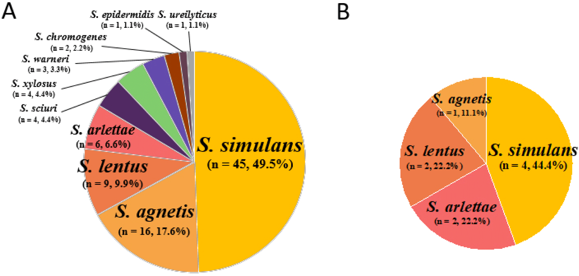
During the study period, 100 NAS isolates of 10 different species were obtained from 270 swab samples (37.04%, 100/270) collected from six different poultry slaughterhouses (Fig. 1A and B). Out of the 100 NAS isolates, 91 NAS isolates were obtained from chicken carcasses, and nine were isolated from the slaughterhouse environments. Based on their ability to produce coagulase, these NAS isolates were divided into coagulase-variable (CoVS; n=19 isolates) and CoNS (n=81; Table 1). While the 19 CoVS isolates comprised only two species of NAS, S. agnetis (n=17) and S. chromogenes (n=2), the 81 CoNS isolates had eight different Staphylococcus species: S. simulans (n=49), S. lentus (n=11), S. arlettae (n=8), S. xylosus (n=4), Staphylococcus sciuri (n=4), Staphylococcus warneri (n=3), S. epidermidis (n=1), and Staphylococcus urealyticus (n=1). The two most prevalent NAS species isolated from the chicken carcasses were S. simulans (49.5%, 45/91 isolates) and S. agnetis (17.6%, 17/91 isolates; Fig. 1A). Similarly, the nine NAS isolates from the facility environments were S. simulans (n=4 isolates), S. arlettae (n=2), S. lentus (n=2), and S. agnetis (n=1; Fig. 1B).

kosfa-45-3-744-g1
Fig. 1. Species distribution of non-aureus staphylococci (NAS) isolated from chicken carcasses (A) and slaughterhouse environments (B) in Korea. In total, 100 NAS isolates from 10 different species were identified in poultry slaughterhouses in Korea.
Download Original Figure
Table 1. Profiles of NAS and SCCmec types of methicillin-resistant NAS strains isolated from poultry slaughterhouses
NAS (n=isolates) mecA positive (%) mec gene ccr gene SCCmec type
CoVS (19)
Staphylococcus agnetis (17) - - - -
Staphylococcus chromogenes (2) - - - -
CoNS (81)
Staphylococcus arlettae (8) - - - -
Staphylococcus epidermidis (1) 1 (100) B A2B2 SCCmec IV
Staphylococcus lentus (11) 6 (54.5) Multi - NT
Multi -
Multi -
Multi -
Multi -
Multi Multi
Staphylococcus simulans (49) 8 (16.3) B A1B6 NT
B A1B1 SCCmec I
B - NT
B A1B1 SCCmec I
B - NT
- A1B6 NT
E - NT
A A1B1 NT
Staphylococcus sciuri (4) - - - -
Staphylococcus urealyticus (1) - - - -
Staphylococcus warneri (3) - - - -
Staphylococcus xylosus (4) - - - -

NAS, non-aureus staphylococci; SCCmec, staphylococcal cassette chromosome mec; ccr, chromosomal cassette recombinase; CoVS, coagulase-variable staphylococci; CoNS, coagulase-negative staphylococci; NT, non-typeable.

Download Excel Table
Detection of mecA and staphylococcal cassette chromosome mec types of methicillin-resistant non-aureus staphylococci

PCR analysis revealed that none of the CoVS isolates were positive for mecA gene (Table 1). Among the 81 CoNS isolates, 15 (18.5%) were positive for mecA: S. simulans (n=8), S. lentus (n=6), and S. epidermidis (n=1). Furthermore, SCCmec element typing of the 15 mecA-positive NAS isolates revealed that eight S. simulans isolates had either non-typeable SCCmec (n=6) or SCCmec I (n=2). In addition, all six methicillin-resistant S. lentus strains contained non-typeable SCCmec elements. One methicillin-resistant S. epidermis isolate from a chicken carcass sample possessed SCCmec IV.

Antimicrobial resistance profiles of the non-aureus staphylococci isolates

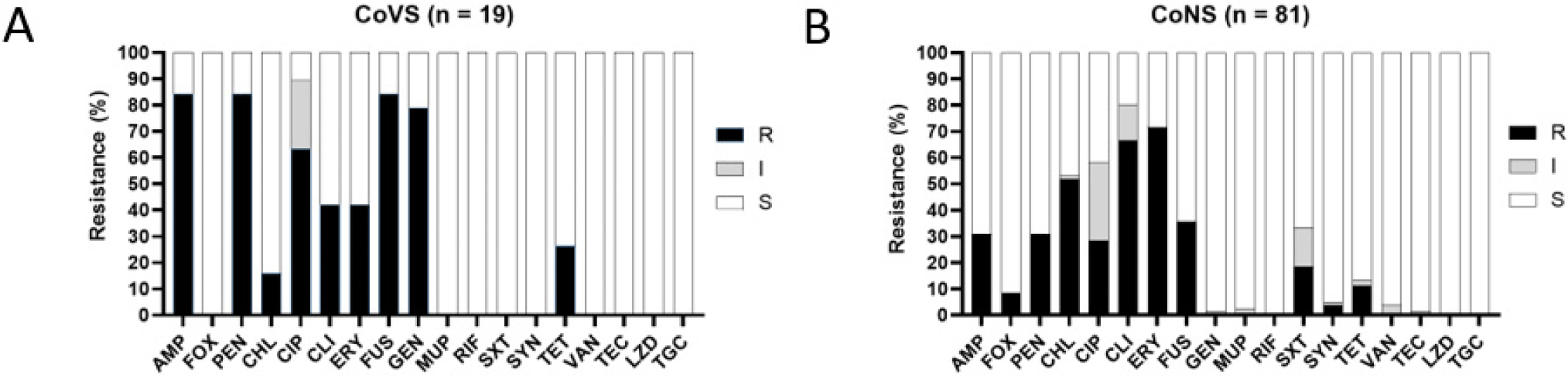
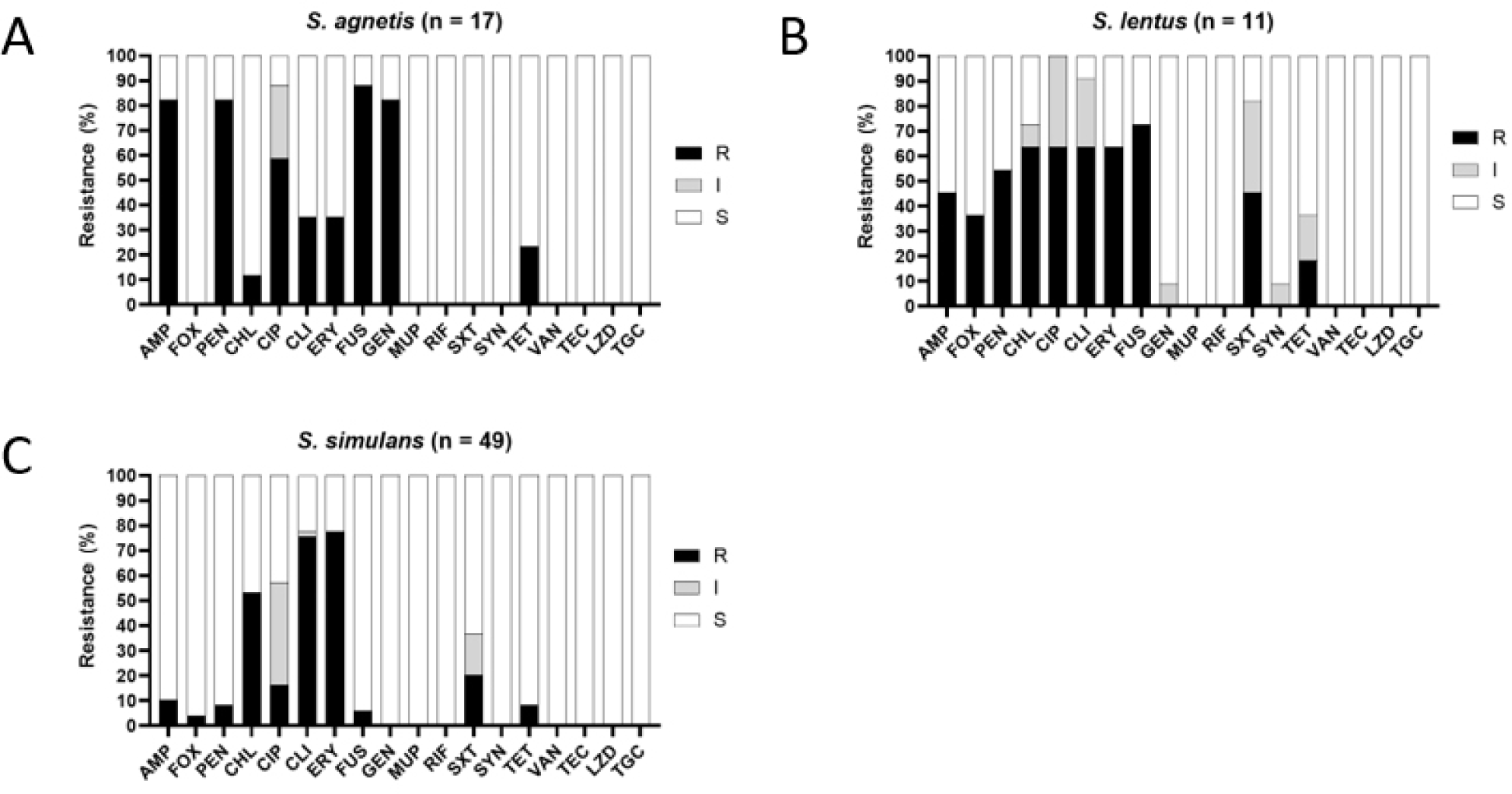
Compared with the other antibiotics, the CoVS isolates exhibited higher resistance to FUS (84.2%, 16/19 isolates), PEN (84.2%, 16/19 isolates), AMP (84.2%, 16/19), GEN (78.9%, 15/19), and CIP (63.2%, 12/19; Table 2 and Fig. 2). Although a higher resistance level to these antibiotics was also observed in CoNS isolates, CoNS exhibited lower resistance rates to the five antibiotics compared with CoVS isolates. In addition, CoVS isolates exhibited higher levels of multidrug resistance (MDR) phenotypes (resistant to three or more classes/subclasses of antimicrobial agents) than CoNS isolates (84.2% vs. 63.0%, respectively). When the AMR of the dominant CoVS species (S. agnetis) and the two major species of CoNS (S. simulans and S. lentus) were compared, S. agnetis (82.4%) and S. lentus (90.9%) exhibited significantly higher degrees of MDR than S. simulans (57.1%; Table 2). Fig. 3 illustrates the heterogeneity of the AMR profiles within the three major species of NAS isolates, S. simulans, S. agnetis, and S. lentus. S. agnetis isolates tended to show high resistance to AMP, GEN, PEN, CIP, and FUS (Fig. 3A). Interestingly, S. lentus, and S. simulans isolates exhibited rather distinct AMR profiles (Fig. 3B and C). However, both S. lentus and S. simulans displayed comparatively high levels of resistance to CHL, CIP, CLI, and ERY. Notably, S. agnetis and S. lentus isolates exhibited higher levels of CIP resistance (61.1% and 63.6%, respectively) than S. simulans isolates. Antimicrobial resistance profiles of each strain are shown in Supplementary Table S2.

Table 2. Antimicrobial resistance profiles of NAS strains isolated from poultry slaughterhouses
NAS (n=isolates) Number of antimicrobial resistance (%)
AMP FOX PEN CHL CIP CLI ERY FUS GEN MUP RIF SXT SYN TET MDR
CoVS (19)
Staphylococcus agnetis (17) 14 (83.3) 0 14 (83.3) 2 (16.7) 10 (61.1) 6 (38.9) 6 (38.9) 15 (88.2) 14 (77.8) 0 0 0 0 4 (27.8) 14 (82.4)
Staphylococcus chromogenes (2) 2 (100) 0 2 (100) 1 (50) 2 (100) 2 (100) 2 (100) 1 (50) 1 (50) 0 0 0 0 1 (50) 2 (100)
CoVS total 16 (84.2) 0 16 (84.2) 3 (15.8) 12 (63.2) 8 (42.1) 8 (42.1) 16 (84.2) 15 (78.9) 0 0 0 0 5 (26.3) 16 (84.2)
CoNS (81)
Staphylococcus arlettae (8) 8 (100) 0 8 (100) 7 (85.7) 7 (85.7) 6 (75) 8 (100) 8 (100) 0 0 0 0 0 3 (37.5) 8 (100)
Staphylococcus epidermidis (1) 1 (100) 1 (100) 1 (100) 0 0 0 1 (100) 1 (100) 0 0 0 0 0 0 1 (100)
Staphylococcus lentus (11) 6 (54.5) 4 (36.4) 6 (54.5) 7 (63.6) 7 (63.6) 7 (63.6) 7 (63.6) 8 (72.7) 0 0 0 5 (45.5) 0 2 (18.2) 10 (90.9)
Staphylococcus sciuri (4) 2 (50) 0 2 (50) 1 (25) 0 0 0 4 (100) 0 0 0 0 0 0 0
Staphylococcus simulans (49) 5 (10.2) 2 (4.1) 4 (8.2) 26 (53.1) 8 (16.3) 37 (75.5) 38 (77.6) 3 (6.1) 0 0 0 10 (20.4) 0 4 (8.2) 28 (57.1)
Staphylococcus urealyticus (1) 0 0 0 1 (100) 0 1 (100) 1 (100) 1 (100) 0 0 0 0 0 0 1 (100)
Staphylococcus warneri (3) 0 0 0 0 0 3 (100) 3 (100) 0 0 0 0 0 3 (100) 0 3 (100)
Staphylococcus xylosus (4) 3 (75) 0 4 (100) 0 0 0 0 4 (100) 0 0 0 0 0 0 0
CoNS total 25 (30.9) 7 (8.6) 25 (30.9) 42 (51.9) 23 (28.4) 54 (66.7) 58 (71.6) 29 (35.8) 0 0 0 15 (18.5) 3 (3.7) 9 (11.1) 51 (63)
Total (100) 41 (41) 7 (7) 41 (41) 45 (45) 35 (35) 62 (62) 66 (66) 45 (45) 15 (15) 0 0 15 (15) 3 (3) 14 (14) 67 (67)

NAS, non-aureus staphylococci; AMP, ampicillin; FOX, cefoxitin; PEN, penicillin; CHL, chloramphenicol; CIP, ciprofloxacin; CLI, clindamycin; ERY, erythromycin; FUS, fusidic acid; GEN, gentamycin; MUP, mupirocin; RIF, rifampin; SXT, trimethoprim-sulfamethoxazole; SYN, quinupristin- dalfopristin; TET, tetracycline; MDR, multi-drug resistance; CoVS, coagulase-variable staphylococci; CoNS, coagulase-negative staphylococci.

Download Excel Table
kosfa-45-3-744-g2
Fig. 2. Antimicrobial resistance patterns of the non-aureus staphylococci (NAS) isolates collected from the slaughterhouses in Korea. Antimicrobial resistance phenotypes of coagulase-variable staphylococci (CoVS; A) and coagulase-negative staphylococci (CoNS; B) isolates. R, resistant; I, intermediate; S, susceptible; AMP, ampicillin; FOX, cefoxitin; PEN, penicillin; CHL, chloramphenicol; CIP, ciprofloxacin; CLI, clindamycin; ERY, erythromycin; FUS, fusidic acid; GEN, gentamicin; MUP, mupirocin; RIF, rifampicin; SXT, trimethoprim-sulfamethoxazole; SYN, quinupristin-dalfopristin; TET, tetracycline; VAN, vancomycin; TEC, teicoplanin; LZD, linezolid; TGC, tigecycline.
Download Original Figure
kosfa-45-3-744-g3
Fig. 3. Antimicrobial resistance patterns of the major non-aureus staphylococci (NAS) collected from poultry slaughterhouses. Antimicrobial resistance profiles of Staphylococcus agnetis (A), Staphylococcus lentus (B), and Staphylococcus simulans (C) isolates. R, resistant; I, intermediate; S, susceptible; AMP, ampicillin; FOX, cefoxitin; PEN, penicillin; CHL, chloramphenicol; CIP, ciprofloxacin; CLI, clindamycin; ERY, erythromycin; FUS, fusidic acid; GEN, gentamicin; MUP, mupirocin; RIF, rifampicin; SXT, trimethoprim-sulfamethoxazole; SYN, quinupristin-dalfopristin; TET, tetracycline; VAN, vancomycin; TEC, teicoplanin; LZD, linezolid; TGC, tigecycline.
Download Original Figure
Mutations in the quinolone-resistance determining regions of fluoroquinolone-resistant non-aureus staphylococci isolates

Among the 100 NAS isolates, 12 CoVS (10 S. agnetis and two S. chromogenes) and 22 CoNS (eight S. simulans, seven S. arlettae, and seven S. lentus) isolates exhibited resistance to CIP (Table 3). Sequencing analysis of QRDRs within gyrA, gyrB, parC, and parE revealed that mutations at codon 84 in gyrA (S84L and D84N) and codon 80 in parC (S80L, T80I, T80R, S80F, and S80Y) were most frequently associated with the CIP-resistant phenotype of the NAS isolates (Table 3). Unlike point mutations in gyrA and parC, a high degree of mutation heterogeneity was observed in gyrB and parE within and between different NAS species (Table 3). Nevertheless, none of the FQ-resistant S. lentus (n=7) isolates carried point mutations in gyrB. Similarly, none of the FQ-resistant S. agnetis isolates harbored point mutations in parE.

Table 3. Point mutations within QRDRs of gyrA, gyrB, parC, and parE genes associated with quinolone resistance in the study strains
NAS species No. of FQ-resistant isolates (%) Mutations in QRDRs Total Zone of inhibition (mm)
gyrA gyrB parC parE
CoVS Staphylococcus agnetis (17) 10 (58.8) S84L - S80L - 6 12.8±1.2
S84L D562Y S80L - 2 13.5±2.1
S84L F522V, K550R, D562Y S80L - 1 13
S84L F522V, E560Q, D562Y, E566Q, E597D S80L - 1 13
Staphylococcus chromogenes (2) 2 (100) S84L I359V, G375A, I379V, T514K H77L, G78A, S80L T348P, D356E, S357A, S360A, D565T 1 6
S84L, I131L H511I, T514N S80L, D84Y K568N 1 13
CoNS Staphylococcus arlettae (8) 7 (87.5) S84L - T80I - 3 6
- - T80I - 1 6
S84L - T80I A531Q 1 6
S84L T80I A360S 1 6
S84L K554N, R561Q, K565Q, K567T T80I K375T, V382G, K383E, R395G, R406Q, K407E, G413S, A418P 1 6
Staphylococcus lentus (11) 7 (63.6) S84L, T172A - S80L - 2 6
S84L, T172A - S80L Y497T 3 6
S84L, A162S, T172A - - Y497T 1 10
S84L, A162S, T172A - S80L Y497T 1 9
Staphylococcus simulans (49) 8 (16.3) S84L, A173S - F74Y, S80F - 1 14
S84L, A173S - S80Y, D84N - 1 6
S84L, A173S A512R S80Y, D84N - 1 6
S84L, A173S A512P S80F, D84N - 1 6
S84L, A173S - S80Y, D84N L377I 1 6
S84L, A173S - S80F, D84N N498K, R499S 1 6
S84L, A132S, A173S E489G, S494T S80I V359I, F365Y, E467D 2 6

QRDRs, quinolone resistance determining regions; NAS, non-aureus staphylococci; FQ, fluoroquinolone; CoVS, coagulase-variable staphylococci; CoNS, coagulase-negative staphylococci.

Download Excel Table
Detection of staphylococcal enterotoxin and toxic shock syndrome toxin-1 genes in non-aureus staphylococci isolates

Among the 100 NAS isolates, only two CoNS isolates (one S. simulans and one S. xylosus) from chicken carcasses were positive for TSST-1 gene. None of the 19 CoVS isolates were positive for the five classical SE genes and tst1.

Discussion

Many recent studies have revealed that NAS are important reservoirs of AMR and enterotoxin genes, which can be transmitted to other pathogenic bacteria, including more pathogenic strains of S. aureus. In our previous research, we suggested that NAS in retail chicken meat can play an important role in the transmission of AMR and enterotoxin genes (Lee et al., 2020). However, not many studies have investigated the species profiles of antimicrobial-resistant NAS and their significance in food-related public health.

In the present study, we investigated NAS species distribution and their AMR profiles in chicken carcasses, facility environments, and poultry slaughterhouse workers in Korea. Similar to the findings of our previous studies on NAS profiles in broiler farms (Park et al., 2023) and retail chicken meat (Lee et al., 2020), the prevalence of CoNS (81.0%, 81/100) was considerably higher than that of CoVS (19.0%, 19/100; Table 1). A higher prevalence of CoNS compared with CoVS has also been reported in both healthy broilers and broilers with signs of illness from other countries. In the present study, S. simulans (49.0%, 49/100) and S. agnetis (17.0%, 17/100) were the most frequently identified NAS species in poultry slaughterhouses; this is consistent with the previously reported high prevalence of S. agnetis, S. simulans, S. haemolyticus, and S. xylosus in broiler farms (Park et al., 2023). Sample origin did not affect the species distribution of NAS isolates in chicken carcasses and facility environments in poultry slaughterhouses (Figs. 1A and B). The four major NAS species, S. simulans, S. agnetis, S. lentus, and S. arlettae, were commonly identified in both chicken carcasses and facility environments. These results suggest that broiler-associated NAS species (Park et al., 2023), both CoVS and CoNS species are potential sources of contaminants in chicken carcasses and facility environments in poultry slaughterhouses.

Approximately 20% of all the antibiotics sold in the livestock industry, particularly FQs, β-lactams, and TETs, are consumed in poultry farming in Korea (Lim et al., 2014). Thus, recent studies on livestock-associated antimicrobial-resistant NAS, particularly methicillin-resistant NAS isolates, have raised significant concerns regarding the transmission of resistance genes to more pathogenic bacteria in the food production system. In a previous study in Switzerland (Huber et al., 2011), researchers reported that 52.8% (38/72) of mecA-positive NAS isolates, i.e., S. lentus (n=30), S. sciuri (n=6), and S. epidermidis (n=2), were obtained from chicken carcasses. In the present study, mecA-positive NAS isolates (15.0%, 15/100) were present at relatively low levels in poultry slaughterhouses. As summarized in Table 1, eight mecA-positive but FOX-susceptible NAS isolates (two S. lentus and six S. simulans) carrying non-typeable SCCmec elements were identified. Recent studies revealed the mecA-positive but FOX-susceptible phenotype of staphylococci (Goering et al., 2019; Ho et al., 2020). This discrepancy could be owing to the altered expression of mecA because of dysregulation in mecR1 (encoding the inducer protein MecR1), mecI (encoding the repressor protein MecI), or bla regulatory system (blaR1-blaI; Liu et al., 2016). In addition, a single-nucleotide insertion within mecA, resulting in the premature termination of mecA expression, has been identified in S. aureus strains (Kime et al., 2019).

Largely correlating with the sales amounts of antibiotics in the poultry industry in Korea, relatively high levels of resistance to β-lactams, GEN, TET, and CIP were identified in NAS isolates, particularly in CoVS isolates. This suggests that antibiotic selective pressure affect the prevalence of antimicrobial-resistant NAS isolates in poultry slaughterhouses (Table 2). Furthermore, the high prevalence of FUS resistance in some of NAS isolates, particularly S. agnetis, S. arlettae, S. lentus, and S. xylosus, may be because of intrinsic FUS resistance owing to the frequent carriage of fusD and fusF (Hung et al., 2015). Notably, the high prevalence of NAS isolates with MDR phenotypes to the antibiotics extensively consumed in poultry farms suggests that antibiotic selective pressure facilitates the proliferation of antimicrobial-resistant NAS in broiler farms, and thus, its transmission to chicken carcasses in slaughterhouses.

The World Health Organization has categorized FQs as the highest-priority critically important antibiotics (Scott et al., 2019). However, FQ-resistant S. aureus and NAS strains have been frequently isolated in livestock farms, particularly poultry farms, in Korea. In this study, 34 FQ-resistant NAS isolates (12 CoVS and 22 CoNS) were identified to harbor multiple point mutations in the QRDRs of DNA gyrase and topoisomerase IV, except for one S. arlettae isolate carrying a single point mutation in parC (Table 3). Consistent with the findings of previous studies of FQ-resistant staphylococci (Takahashi et al., 1998; Wang et al., 2013), all 34 FQ-resistant NAS isolates possessed point mutations in gyrA (codon 84) or parC (codon 80). In contrast to the mutations in gyrA and parC, which are frequently concentrated in codons 84 and 80, respectively, point mutations in gyrB and parE were identified at various locations in QRDRs. Moreover, previous studies on FQ-resistant staphylococci (Nakaminami et al., 2014; Yamada et al., 2008) revealed a high degree of heterogeneity in the point mutations in gyrB and parE. Although major mutations, including S84L at gyrA, S80L at parC, and D84N at parC, were previously known to be associated with FQ resistance (Li et al., 1998; Takahashi et al., 1998; Vila et al., 1997), the precise role of other minor mutations in FQ resistance should be elucidated in the future research.

SEs produced by coagulase-positive S. aureus are a major cause of SFP (Argudín et al., 2010; Fisher et al., 2018). However, many recent studies have indicated that NAS isolates, including those from livestock and food products, carry SE genes and are potentially enterotoxigenic (Mekhloufi et al., 2021; Podkowik et al., 2013). In this study, our results demonstrate that CoVS isolates from poultry slaughterhouses do not harbor the classical SE nor tst-1 genes. Although previous studies have revealed the highest prevalence of sec and tst1 in CoNS strains (Hájek, 1978; Valle et al., 1990), only two tst1-positive (one S. simulans and one S. xylosus) NAS isolates were detected in the present study. Since SEA and TSST-1 were first detected in CoNS strains isolated from patients with toxic shock syndromes (Crass and Bergdoll, 1986), potential enterotoxigenic or pathogenic NAS strains carrying SE genes have been detected in humans (Achek et al., 2018; Banaszkiewicz et al., 2022), livestock (Ruiz-Ripa et al., 2020; Ünal and Çinar, 2012), and ready-to-eat foods (Chajęcka-Wierzchowska et al., 2020; Crass and Bergdoll, 1986; Cunha et al., 2006; Rall et al., 2010; Zell et al., 2008). Despite the proposed instability of SE genes in CoNS isolates (Banaszkiewicz et al., 2022), the carriage of SE and TSST-1 genes in NAS isolates from poultry slaughterhouses should be monitored as a significant threat to food safety.

Conclusion

In summary, this is the first report on the species diversity, AMR patterns, and genetic characteristics of methicillin- and FQ-resistant NAS isolates obtained from poultry slaughterhouses in Korea. Our findings suggest that (i) the overall species distribution of NAS isolates in poultry slaughterhouses is similar to that in broiler farms (Park et al., 2023) in Korea; (ii) MDR phenotypes with relatively high resistance levels of resistance to PEN, AMP, GEN, CIP, and TET are observed in NAS isolates; (iii) FQ-resistance in NAS isolates is caused by point mutations occurring at specific locations (gyrA and parC) or rather various locations (gyrB and parE) of QRDRs; and (iv) the carriage of tst1 only in CoNS isolates, indicating the extremely low prevalence of the TSST-1 and SE genes in NAS isolates. In addition, this study underscores a potential role of poultry slaughterhouses in transmission of antimicrobial-resistant NAS strains to human food chains.

To combat AMR in poultry industry, a multisectoral approach, including poultry farms, slaughterhouses, and human workers, is necessary. Improper use of antibiotics and indiscriminate prescription of antibiotics should be avoided to prevent occurrence and amplification of antimicrobial-resistant bacteria in poultry farms. Furthermore, importance of hygiene measures in slaughterhouses should be emphasized along with a multisectoral surveillance networks to better detect and mitigate spread of AMR.

Supplemenrary Materials

Supplementary Materials

kosfa-45-3-744-suppl1.pdf

Conflicts of Interest

The authors declare no potential conflicts of interest.

Acknowledgements

This study was supported by funding from the Research of Korea Centers for Disease Control and Prevention (Projects No. 2017NER54060 & 2021ER220100).

Author Contributions

Conceptualization: Lee KJ, Yang SJ. Data curation: Lim JH, Yang SJ. Formal analysis: Lim JH, Park JH, Lee GY, Lee JB. Methodology: Lim JH, Park JH, Lee GY. Investigation: Lim JH, Park JH, Yang SJ. Writing - original draft: Lim JH, Lee JB, Lee KJ, Yang SJ. Writing - review & editing: Lim JH, Park JH, Lee GY, Lee JB, Lee KJ, Yang SJ.

Ethics Approval

This article does not require IRB/IACUC approval because there are no human and animal participants.

References

1.

Abdalrahman LS, Stanley A, Wells H, Fakhr MK. 2015; Isolation, virulence, and antimicrobial resistance of methicillin-resistant Staphylococcus aureus (MRSA) and methicillin sensitive Staphylococcus aureus (MSSA) strains from Oklahoma retail poultry meats. Int J Environ Res Public Health. 12:6148-6161

2.

Achek R, Zafer C, Oumouna M, Mahdi A, Hamdi TM. 2018; Occurrence of enterotoxins, exfoliative toxins and toxic shock syndrome toxin-1 genes in Staphylococcus aureus and CoNS isolated from clinical and food samples in Algeria. Hum Vet Med Int J Bioflux Soc. 10:85-92.

3.

Adkins PRF, Dufour S, Spain JN, Calcutt MJ, Reilly TJ, Stewart GC, Middleton JR. 2018; Molecular characterization of non-aureusStaphylococcus spp. from heifer intramammary infections and body sites. J Dairy Sci. 101:5388-5403

4.

Alibayov B, Baba-Moussa L, Sina H, Zdeňková K, Demnerová K. 2014; Staphylococcus aureus mobile genetic elements. Mol Biol Rep. 41:5005-5018

5.

Apata DF. 2009; Antibiotic resistance in poultry. Int J Poult Sci. 8:404-408

6.

Argudín MÁ, Mendoza MC, Rodicio MR. 2010; Food poisoning and Staphylococcus aureus enterotoxins. Toxins. 2:1751-1773

7.

Banaszkiewicz S, Walecka-Zacharska E, Schubert J, Tabiś A, Król J, Stefaniak T, Węsierska E, Bania J. 2022; Staphylococcal enterotoxin genes in coagulase-negative staphylococci: Stability, expression, and genomic context. Int J Mol Sci. 23:2560

8.

Becker K, Heilmann C, Peters G. 2014; Coagulase-negative staphylococci. Clin Microbiol Rev. 27:870-926

9.

Bhargava K, Zhang Y. 2012; Multidrug‐resistant coagulase‐negative staphylococci in food animals. J Appl Microbiol. 113:1027-1036

10.

Casey AL, Lambert PA, Elliott TSJ. 2007; Staphylococci. Int J Antimicrob Agents. 29:S23-S32

11.

Chajęcka-Wierzchowska W, Gajewska J, Wiśniewski P, Zadernowska A. 2020; Enterotoxigenic potential of coagulase-negative staphylococci from ready-to-eat food. Pathogens. 9:734

12.

Clinical and Laboratory Standards Institute [CLSI]. 2015 VET01S: Performance standards for antimicrobial disk and dilution susceptibility tests for bacteria isolated from animals. 3rd edClinical and Laboratory Standards Institute. Wayne, PA, USA: .

13.

Clinical and Laboratory Standards Institute [CLSI]. 2022 M100: Performance standards for antimicrobial susceptibility testing. 32nd edClinical and Laboratory Standards Institute. Wayne, PA, USA: .

14.

Crass BA, Bergdoll MS. 1986; Involvement of coagulase-negative staphylococci in toxic shock syndrome. J Clin Microbiol. 23:43-45

15.

Creţu C, Bondoc I, Obadă MD, Carp-Cărare M. 2011. Determining pathogenicity strains of Salmonella spp. isolated from the carcasses of poultry. Available from: https://www.researchgate.net/publication/323998291_determining_pathogenicity_strains_of_salmonella_spp_isolated_from_the_carcasses_of_poultryAccessed at Apr 2, 2024.

16.

Creţu C, Horhogea C, Rîmbu C, Bondoc I. 2015. Research on the need for use decontamination of poultry carcasses. Available from: https://www.researchgate.net/publication/323998056_Research_on_the_need_for_use_decontamination_of_poultry_carcassesAccessed at Apr 2, 2024.

17.

Creţu C, Obadă MD, Floriştean V, Bondoc I, Carp Cărare M. 2012. Determining pathogenicity strains of Salmonella spp. isolated from the carcasses of poultry. Available from: https://www.researchgate.net/publication/323998291_determining_pathogenicity_strains_of_salmonella_spp_isolated_from_the_carcasses_of_poultryAccessed at Apr 2, 2024.

18.

Cunha MDLRDSD, Peresi E, Calsolari RAO, Araújo JP. 2006; Detection of enterotoxins genes in coagulase-negative staphylococci isolated from foods. Braz J Microbiol. 37:70-74

19.

Dalhoff A. 2012; Global fluoroquinolone resistance epidemiology and implictions for clinical use. Interdiscip Perspect Infect Dis. 2012:976273

20.

Dellaripa PF. 2000; Toxic shock syndrome. J Intensive Care Med. 15:314-320

21.

Diarra MS, Malouin F. 2014; Antibiotics in Canadian poultry productions and anticipated alternatives. Front Microbiol. 5:282

22.

Feßler AT, Kadlec K, Hassel M, Hauschild T, Eidam C, Ehricht R, Monecke S, Schwarz S. 2011; Characterization of methicillin-resistant Staphylococcus aureus isolates from food and food products of poultry origin in Germany. Appl Environ Microb. 77:7151-7157

23.

Fisher EL, Otto M, Cheung GYC. 2018; Basis of virulence in enterotoxin-mediated staphylococcal food poisoning. Front Microbiol. 9:436

24.

Geha DJ, Uhl JR, Gustaferro CA, Persing DH. 1994; Multiplex PCR for identification of methicillin-resistant staphylococci in the clinical laboratory. J Clin Microbiol. 32:1768-1772

25.

Goering RV, Swartzendruber EA, Obradovich AE, Tickler IA, Tenover FC. 2019; Emergence of oxacillin resistance in stealth methicillin-resistant Staphylococcus aureus due to mecA sequence instability. Antimicrob Agents Chemother. 63e00558-19

26.

Hájek V. 1978; Identification of enterotoxigenic staphylococci from sheep and sheep cheese. Appl Environ Microbiol. 35:264-268

27.

Ho PL, Liu MCJ, Tong MK, Fan PM, Tse CWS, Wu AKL, Cheng VCC, Chow KH. 2020; Evaluation of disc diffusion tests and agar screening for predicting mecA-mediated oxacillin resistance in Staphylococcus lugdunensis revealed a cefoxitin-susceptible, mecA-positive S. lugdunensis clonal complex 27 clone. J Glob Antimicrob Resist. 20:260-265

28.

Huber H, Ziegler D, Pflüger V, Vogel G, Zweifel C, Stephan R. 2011; Prevalence and characteristics of methicillin-resistant coagulase-negative staphylococci from livestock, chicken carcasses, bulk tank milk, minced meat, and contact persons. BMC Vet Res. 7:1-7

29.

Hung WC, Chen HJ, Lin YT, Tsai JC, Chen CW, Lu HH, Tseng SP, Jheng YY, Leong KH, Teng LJ. 2015; Skin commensal staphylococci may act as reservoir for fusidic acid resistance genes. PLOS ONE. 10e0143106

30.

Ito H, Yoshida H, Bogaki-Shonai M, Niga T, Hattori H, Nakamura S. 1994; Quinolone resistance mutations in the DNA gyrase gyrA and gyrB genes of Staphylococcus aureus. Antimicrob Agents Chemother. 38:2014-2023

31.

Kime L, Randall CP, Banda FI, Coll F, Wright J, Richardson J, Empel J, Parkhill J, O’Neill AJ. 2019; Transient silencing of antibiotic resistance by mutation represents a significant potential source of unanticipated therapeutic failure.. mBio. 10e01755-19

32.

Kondo Y, Ito T, Ma XX, Watanabe S, Kreiswirth BN, Etienne J, Hiramatsu K. 2007; Combination of multiplex PCRs for staphylococcal cassette chromosome mec type assignment: Rapid identification system for mec, ccr, and major differences in junkyard regions. Antimicrob Agents Chemother. 51:264-274

33.

Ladhani S, Konana OS, Mwarumba S, English MC. 2004; Bacteraemia due to Staphylococcus aureus. Arch Dis Child. 89:568-571

34.

Lee GY, Lee SI, Kim SD, Park JH, Kim GB, Yang SJ. 2022; Clonal distribution and antimicrobial resistance of methicillin-susceptible and -resistant Staphylococcus aureus strains isolated from broiler farms, slaughterhouses, and retail chicken meat. Poult Sci. 101:102070

35.

Lee SI, Kim SD, Park JH, Yang SJ. 2020; Species distribution, antimicrobial resistance, and enterotoxigenicity of non-aureus staphylococci in retail chicken meat. Antibiotics. 9:809

36.

Li Z, Deguchi T, Yasuda M, Kawamura T, Kanematsu E, Nishino Y, Ishihara S, Kawada Y. 1998; Alteration in the gyrA subunit of DNA gyrase and the parC subunit of DNA topoisomerase IV in quinolone-resistant clinical isolates of Staphylococcus epidermidis. Antimicrob Agents Chemother. 42:3293-3295

37.

Lim SK, Lee JE, Lee HS, Nam HM, Moon DC, Jang GC, Park YJ, Jung YG, Jung SC, Wee SH. 2014; Trends in antimicrobial sales for livestock and fisheries in Korea during 2003-2012. Korean J Vet Res. 54:81-86

38.

Lim SK, Nam HM, Park HJ, Lee HS, Choi MJ, Jung SC, Lee JY, Kim YC, Song SW, Wee SH. 2010; Prevalence and characterization of methicillin-resistant Staphylococcus aureus in raw meat in Korea. J Microbiol Biotechnol. 20:775-778.

39.

Liu P, Xue H, Wu Z, Ma J, Zhao X. 2016; Effect of bla regulators on the susceptible phenotype and phenotypic conversion for oxacillin-susceptible mecA-positive staphylococcal isolates. J Antimicrob Chemother. 71:2105-2112

40.

Lowy FD. 1998; Staphylococcus aureus infections. N Engl J Med. 339:520-532

41.

Mekhloufi OA, Chieffi D, Hammoudi A, Bensefia SA, Fanelli F, Fusco V. 2021; Prevalence, enterotoxigenic potential and antimicrobial resistance of Staphylococcus aureus and methicillin-resistant Staphylococcus aureus (MRSA) isolated from Algerian ready to eat foods. Toxins. 13:835

42.

Nakaminami H, Sato-Nakaminami K, Noguchi N. 2014; A novel gyrB mutation in meticillin-resistant Staphylococcus aureus (MRSA) confers a high level of resistance to third-generation quinolones. Int J Antimicrob Agents. 43:478-479

43.

Ng EY, Trucksis M, Hooper DC. 1996; Quinolone resistance mutations in topoisomerase IV: Relationship to the flqA locus and genetic evidence that topoisomerase IV is the primary target and DNA gyrase is the secondary target of fluoroquinolones in Staphylococcus aureus. Antimicrob Agents Chemother. 40:1881-1888

44.

Normanno G, Corrente M, La Salandra G, Dambrosio A, Quaglia NC, Parisi A, Greco G, Bellacicco AL, Virgilio S, Celano GV. 2007; Methicillin-resistant Staphylococcus aureus (MRSA) in foods of animal origin product in Italy. Int J Food Microbiol. 117:219-222

45.

Omoe K, Hu DL, Takahashi-Omoe H, Nakane A, Shinagawa K. 2005; Comprehensive analysis of classical and newly described staphylococcal superantigenic toxin genes in Staphylococcus aureus isolates. FEMS Microbiol Lett. 246:191-198

46.

Osman K, Alvarez-Ordóñez A, Ruiz L, Badr J, ElHofy F, Al-Maary KS, Moussa IMI, Hessain AM, Orabi A, Saad A, Elhadidy M. 2017; Antimicrobial resistance and virulence characterization of Staphylococcus aureus and coagulase-negative staphylococci from imported beef meat. Ann Clin Microbiol Antimicrob. 16:35

47.

Osman K, Badr J, Al-Maary KS, Moussa IMI, Hessain AM, Girah ZMSA, Abo-Shama UH, Orabi A, Saad A. 2016; Prevalence of the antibiotic resistance genes in coagulase-positive- and negative-Staphylococcus in chicken meat retailed to consumers. Front Microbiol. 7:1846

48.

Park JH, Lee GY, Lim JH, Kim GB, Park KT, Yang SJ. 2023; Species profiles and antimicrobial resistance of non-aureus staphylococci isolated from healthy broilers, farm environments, and farm workers. Food Sci Anim Resour. 43:792-804

49.

Pimenta RL, de Melo DA, Bronzato GF, de Salles Souza VR, Holmström TCN, de Oliveira Coelho SM, da Silva Coelho I, de Souza MMS. 2021; Characterization of Staphylococcus spp. isolates and β-lactam resistance in broiler chicken production. Braz J Vet Med. 43e00720

50.

Podkowik M, Bystroń J, Bania J. 2012; Prevalence of antibiotic resistance genes in staphylococci isolated from ready-to-eat meat products. Pol J Vet Sci. 15:233-237

51.

Podkowik M, Park JY, Seo KS, Bystroń J, Bania J. 2013; Enterotoxigenic potential of coagulase-negative staphylococci. Int J Food Microbiol. 163:34-40

52.

Pyzik E, Marek A, Stępien-Pyśniak D, Urban-Chmiel R, Jarosz ŁS, Jagiełło-Podębska I. 2019; Detection of antibiotic resistance and classical enterotoxin genes in coagulase-negative staphylococci isolated from poultry in Poland. J Vet Res. 63:183-190

53.

Rall VLM, Sforcin JM, de Deus MFR, De Sousa DC, Camargo CH, Godinho NC, Galindo LA, Soares TCS, Araújo JP 2010; Polymerase chain reaction detection of enterotoxins genes in coagulase-negative staphylococci isolated from Brazilian minas cheese. Foodborne Pathog Dis. 7:1121-1123

54.

Ray MD, Boundy S, Archer GL. 2016; Transfer of the methicillin resistance genomic island among staphylococci by conjugation. Mol Microbiol. 100:675-685

55.

Ruiz-Ripa L, Gómez P, Alonso CA, Camacho MC, Ramiro Y, de la Puente J, Fernández-Fernández R, Quevedo MÁ, Blanco JM, Báguena G. 2020; Frequency and characterization of antimicrobial resistance and virulence genes of coagulase-negative staphylococci from wild birds in Spain. Detection of tst-carrying S. sciuri isolates. Microorganisms. 8:1317

56.

Schnitt A, Lienen T, Wichmann-Schauer H, Tenhagen BA. 2021; The occurrence of methicillin-resistant non-aureus staphylococci in samples from cows, young stock, and the environment on german dairy farms. J Dairy Sci. 104:4604-4614

57.

Scott HM, Acuff G, Bergeron G, Bourassa MW, Gill J, Graham DW, Kahn LH, Morley PS, Salois MJ, Simjee S, Singer RS, Smith TC, Storrs C, Wittum TE. 2019; Critically important antibiotics: Criteria and approaches for measuring and reducing their use in food animal agriculture. Ann NY Acad Sci. 1441:8-16

58.

Takahashi H, Kikuchi T, Shoji S, Fujimura S, Lutfor AB, Tokue Y, Nukiwa T, Watanabe A. 1998; Characterization of gyrA, gyrB, grlA and grlB mutations in fluoroquinolone-resistant clinical isolates of Staphylococcus aureus. J Antimicrob Chemother. 41:49-57

59.

Ünal N, Çinar OD. 2012; Detection of stapylococcal enterotoxin, methicillin-resistant and panton–valentine leukocidin genes in coagulase-negative staphylococci isolated from cows and ewes with subclinical mastitis. Trop Anim Health Prod. 44:369-375

60.

Valle J, Gomez-Lucia E, Piriz S, Goyache J, Orden J, Vadillo S. 1990; Enterotoxin production by staphylococci isolated from healthy goats. Appl Environ Microbiol. 56:1323-1326

61.

Van Cleef BAGL, Broens EM, Voss A, Huijsdens XW, Züchner L, Van Benthem BHB, Kluytmans JAJW, Mulders MN, Van De Giessen AW. 2010; High prevalence of nasal MRSA carriage in slaughterhouse workers in contact with live pigs in the Netherlands. Epidemiol Infect. 138:756-763

62.

Vila J, Ruiz J, Goñi P, Jimenez de Anta T. 1997; Quinolone-resistance mutations in the topoisomerase IV parC gene of Acinetobacter baumannii. J Antimicrob Chemother. 39:757-762

63.

Wang SC, Wang Y, Shen J, Wu Y, Wu C. 2013; Polymorphic mutation frequencies in clinical isolates of Staphylococcus aureus: The role of weak mutators in the development of fluoroquinolone resistance. FEMS Microbiol Lett. 341:13-17

64.

Wiśniewski P, Gajewska J, Zadernowska A, Chajęcka-Wierzchowska W. 2023; Identification of the enterotoxigenic potential of Staphylococcus spp. from raw milk and raw milk cheeses. Toxins. 16:17

65.

Wuytack A, De Visscher A, Piepers S, Boyen F, Haesebrouck F, De Vliegher S. 2020; Distribution of non-aureus staphylococci from quarter milk, teat apices, and rectal feces of dairy cows, and their virulence potential. J Dairy Sci. 103:10658-10675

66.

Yamada M, Yoshida J, Hatou S, Yoshida T, Minagawa Y. 2008; Mutations in the quinolone resistance determining region in Staphylococcus epidermidis recovered from conjunctiva and their association with susceptibility to various fluoroquinolones. Br J Ophthalmol. 92:848-851

67.

Zell C, Resch M, Rosenstein R, Albrecht T, Hertel C, Götz F. 2008; Characterization of toxin production of coagulase-negative staphylococci isolated from food and starter cultures. Int J Food Microbiol. 127:246-251