


Introduction
Brazil is an important broiler feet exporter to the Asian trade, especially China and Hong Kong, which consider this product as a delicacy in their gastronomy and as collagen supplement for the cosmetic and medicine industry (ApexBrasil, 2018; Munasinghe et al., 2014). Moreover, it is considered in many countries, as well as in Brazil, as the third cut of chicken with the highest economic value (Chen et al., 2016).
Among the main factors for the condemnation of broiler feet are the contact lesions and keratosis. The contact lesions in the tibiotarsal region, also known as hock burn, contact dermatitis and dermatitis are characterized by the presence of blackish or brownish coloration on the skin of the chickens’ tibiotarsal region (Bessei, 2006). Besides the economic losses due to discards, it also impairs the broilers performance and violates the technical recommendation of animal welfare in poultry production, because it results in pain for the bird, evidenced by the slowly locomotion or even reluctance to move (Louton et al., 2020).
It is known that the weight is a risk factor that can be associated with the occurrence of lesions on broiler legs (Louton et al., 2018) and that it will simultaneously acts alongside the bad litter quality and the presence of infectious agents (Thøfner et al., 2019). The litter of the poultry house is used to provide a better life quality for the birds since it contributes to the thermal comfort and avoid direct contact with the floor or tread, preventing the formation of calluses in the animal’s leg and breast. Besides the litter absorbs and incorporates the waste, such as excrete, desquamation, feathers and food and water residues that fall from the feeders and drinking fountains (Avila et al., 2008). According to Heidemann Olsen et al. (2018), the high humidity and high ammonia concentrations originated from the accumulated faecal matter result in ammonia gas liberation that causes chemical burns and weakening of the dermis. In addition, the humidity makes the external dermis softens, facilitating the entry and proliferation of microorganisms.
Although a visual classification system for contact lesion exists (Michel et al., 2012), a histopathological validation for this assessment scheme was just recently described (Louton et al., 2020). Besides, until now, only the correspondence between the macroscopic aspects of the lesions and their histopathological characteristic was demonstrated with no correlation between their microbiological content. Nevertheless, since no standard visual method was developed to compare this kind of lesion, this study aimed to present a fast and practical visual grade to be used in the industry and validated with the histological findings and microbiological quantification in the tibiotarsal region in broiler carcasses.
Materials and Methods
This study was conducted in a commercial slaughterhouse located in the Sao Paulo state, Brazil, with a slaughter capacity of 120.000 birds/day. The birds came from 14 producers of the Northern regions of the Sao Paulo state. The bird batches were of the Cobb and Ross linages, with a mean of 42 days of age and 2.8 kg of weight at the slaughter.
For this study, 112 broiler carcasses were obtained at the post-mortem inspection line (pre-inspection) with or without visual characteristics of contact lesion at the tibiotarsal region. Then, carcasses were removed from the inspection line and with the aid of a disinfect knife in alcohol 70%, the leg was removed by disarticulating the tibiofemoral region, followed by the disarticulation and removal of the metatarsophalangeal region. Each sample was constituted by a single joint, having collected 28 articulations for each evaluated grade (n=28), which were packed either in sterile bag under refrigeration or flasks containing 10% formaldehyde and, thus, sent to the laboratory for analysis.
For evaluation and visual classification of the tibiotarsal joint, to attribute a macroscopic score of the lesions, was carried out over a period of six months and the lesions were divided into four different groups: a control group (CG) and grades 1 (G1), 2 (G2) and 3 (G3). It was used a total of 942 (CG=234, G1=236, G2=235, and G3=237) pictures of tibiotarsal region observed in the post-mortem inspection line to perform macroscopic classification. The grading methods was based on the principals described by Welfare-Quality® (Welfare Quality Consortium, 2009) and Louton et al. (2020), utilizing fewer lesion grades (three instead of four) that were created based on characteristics such as intensity and color of the contact lesion in the tibiotarsal region and the presence or absence of scarification.
The samples harvested for histopathological examination were placed in flasks containing 10% formalin and sent to the “Centro de Diagnóstico de Sanidade Animal” (CEDISA, Concordia, SC, Brazil). The histological sections were stained with hematoxylin and eosin for evaluation of the epidermis, dermis and hypodermis (subcutaneous) of the tibiotarsal region.
The microbiological analysis were performed at the “Laudo Laboratório Avícola Uberlândia Ltda” (LAUDO, Uberlandia, MG, Brazil). From the refrigerated samples for microbiological count, sterile swabs were scraped on a 5 cm2 area of the articular surface. After sampling, the swabs were placed in tubes containing 5 mL of 1% buffered peptone water solution (Neogen, Lansing, MI, USA). After sampling, E. coli, Staphylococcus spp. and sulphite-reducing clostridia were quantified using 3MTM PetrifilmTM Plates (3M do Brazil, Sumaré, Brazil), and Streptococcus spp. used Blood agar with 5% sheep blood plates (Laborclin, Pinhais, PR, Brazil) as described by Chadfield et al. (2004).
Microbiological data were subjected to statistical analysis using the R-Project for Statistical Computing (Wilson and Norden, 2015) and Python for Data Analysis software (Tobergte and Curtis, 2013). Microbiological counts were logarithmic transformed and after not passing the normality test (Shapiro-Wilk), the results were evaluated with a non-parametric Kruskal-Wallis test and statistical significance of the groups were evaluated by the paired sample Wilcoxon test and considered statistically significant when the p-value was lower than 5% (p<0.05).
Results and Discussion
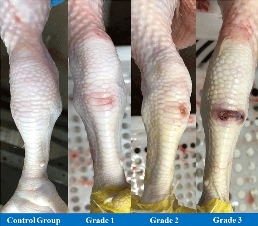
Considering the principals described by Welfare-Quality® (Welfare Quality Consortium, 2009) and Louton et al. (2020) and the characteristics about the intensity and coloration of the contact lesions at the tibiotarsal region, the lesions were allocated in four different grades: The CG the articulation without visual alteration; Grade 1 (G1), refers to the joint with mild scarification and pink or reddish color (erythema); Grade 2 (G2), refers to the joint with moderate scarification, crust formation and brownish coloration; Grade 3 (G3) refers to the articulation with scarification of severe intensity, presence of crust, ulceration and/or brownish and/or purplish and/or blackish coloration (Fig. 1).
The classification of contact injuries prove to be widely diversified, both nationally and internationally. Some of them focus on the size of the lesion (Arnould et al., 2009; Pagazaurtundua and Warriss, 2006a; Pagazaurtundua and Warriss, 2006b; Welfare Quality Consortium, 2009) whereas others combine size and depth (Allain et al., 2009; Ekstrand et al., 1998; Kaukonen et al., 2016; Kjaer et al., 2006) in different categories. Therefore, the differences of this varieties of assessment systems may not well elucidated and has been a challenge for both scientific point of view and the quality of surveillance systems (Michel et al., 2012; Riber et al., 2020). Thus, it was suggested herein a model for these gross lesions classifications to further be used in accordance with histological and microbiological analysis.
In the histopathological characterization was described that in the CG articulation was not observed noteworthy lesions. In the G1 and G2 groups, it was observed histological alterations only in the superficial regions of the skin, whereas in G1, there were a moderate proliferation of subcutaneous connective tissue, with no signs of inflammation. Moreover, in G2 was observed a moderate proliferation of connective tissue, with a mild ulceration focus of the skin and formation of a keratin crust and degenerated inflammatory cells, considered as a morphological diagnose of mild focal necrotic dermatitis.
In G3 were observed ulceration in the epidermis and dermis, with the presence of crusts of necrotic tissue, keratin, degenerated inflammatory cells and clumps of bacteria in the form of cocci. In the subcutaneous (hypoderm), it was observed proliferation of connective tissue, with deposition of fibrin lump and, mild presence of hemorrhage and inflammatory infiltrate, with the heterophile predominance. These characteristics are considered a morphological find of focally extensive necrotic dermatitis. The inflammation severity of the lesions increased with the crescent macroscopic score, in which the visually more severe and deeper lesions, also presented more accentuated histological findings, especially considering the presence of inflammatory cell and remnants of inflammation (proliferation of connective tissue and presence of fibrin; Martins et al., 2016).
The microbiological quantification results, described in Table 1, corroborates with the histopathological characterization, in which no inflammatory reaction of bacterial origin was observed in samples from CG and G1 and G2 and bacterial cocci clumps in G3 samples. In the evaluated samples was not evidenced the presence of sulfite-reducing clostridia, and the counts of E. coli, Staphylococcus spp. and Streptococcus spp. of the CG and grade 1 and 2 were within the acceptance threshold required by Brazil and importing trades such as the USA, China, Eurasian Economic Union, Saudi Arabia, South Africa, and others [Agri-Food and Veterinary Authority of Singapore (AVA), 2000; European Commission (EC), 2005; GB National Standards of People's Republic of China, 2014; GCC Standardization Organization (GSO), 2014; IBNORCA, 2019; Ministry of Agriculture, Livestock and Supply, 2017; Ministry of Agriculture, Livestock and Supply, 2019]. By statistical tests, when comparing the results of the CG with the other grades, it was not observed significant differences (p>0.05), except in G3, which differed from the CG and other grades.
Although there is an extensive repertoire of information about the predisposing factors for the occurrence of contact injuries in tibiotarsal joints of broiler, especially those related to bedding quality, nutrition, and management, little is known about the microbiological factors associated with these conditions and that could be considered inadequate (Heidemann Olsen et al., 2018). Chadfield et al. (2004) suggested that E. coli, Staphylococcus spp. and Streptococcus spp. are opportunistic bacteria that can prevenient from the bird's own organism, which would explain the similarity (p>0.05) in bacterial counts observed for G1 and G2 when compared to the CG.
In the case of mild lesions (G1), in which the skin was preserved intact and in the moderate lesions (G2), where only the superficial skin layer was compromised, there was no microbial invasion into the tissues, which Allain et al. (2009) characterize them as “chemical burns” that are the results of the high amount of humidity of the litter and/or high concentration of ammonia in the fecal matter. In contrast, the accentuated skin deterioration process allow for the opportunistic microorganism to invade the subcutaneous tissue (Nagase et al., 2002). That fact was observed in the present study in lesions of G3, in which there was the presence of ulceration and increased bacterial counts, as described by Martins et al. (2016) who observed the presence of areas of necrosis and the presence of inflammatory infiltrate in severe contact lesions.
Thus, the classification of lesions into three scores of hock dermatitis proposed in this work by means of macroscopic, histopathological and microbiological findings provided a simple, practical and scientific-based way for its use both in the poultry industry, as well as in poultry farms in Brazil. Such grouping allows for easier identification, in addition to enabling better characterization of the evolution of changes in the animals, a fact evidenced both by histopathological findings (presence of connective tissue, inflammatory infiltrate, ulceration and the formation of keratin crust) and by microbiological findings (increasing microorganisms quantification).
Conclusion
The visual standard proposed in this work, which was correlated and confirmed by the histopathological and microbiological characterization, allows a faster and precise ascertainment of the degrees of contact lesions in tibiotarsal joints of broilers. Furthermore, it is noteworthy that grades 1 and 2 alterations are not due to an inflammatory process caused by pathogens and, therefore, do not represent a public health concern.