ARTICLE

Muscle Fiber Typing in Bovine and Porcine Skeletal Muscles Using Immunofluorescence with Monoclonal Antibodies Specific to Myosin Heavy Chain Isoforms

Sumin Song1https://orcid.org/0000-0001-7115-2253, Chi-Hoon Ahn2https://orcid.org/0000-0003-3643-0566, Gap-Don Kim1,2,*https://orcid.org/0000-0001-5870-8990
Author Information & Copyright
1Graduate School of International Agricultural Technology, Seoul National University, Pyeongchang 25354, Korea
2Institutes of Green Bio Science & Technology, Seoul National University, Pyeongchang 25354, Korea
*Corresponding author: Gap-Don Kim, Graduate School of International Agricultural Technology, Seoul National University, Pyeongchang 25354, Korea, Tel: +82-33-339-5778, Fax: +82-33-339-5779, E-mail: gapdonkim@snu.ac.kr

© Korean Society for Food Science of Animal Resources. This is an Open-Access article distributed under the terms of the Creative Commons Attribution Non-Commercial License (http://creativecommons.org/licenses/by-nc/3.0) which permits unrestricted non-commercial use, distribution, and reproduction in any medium, provided the original work is properly cited.

Received: Oct 20, 2019 ; Revised: Dec 11, 2019 ; Accepted: Dec 12, 2019

Published Online: Jan 01, 2020

Abstract

The aim of this study was to optimize staining procedures for muscle fiber typing efficiently and rapidly in bovine and porcine skeletal muscles, such as longissimus thoracis, psoas major, semimembranosus, and semitendinosus muscles. The commercially available monoclonal anti-myosin heavy chain (MHC) antibodies and fluorescent dye-conjugated secondary antibodies were applied to immunofluorescence histology. Two different procedures, such as cocktail and serial staining, were adopted to immunofluo-rescence analysis. In bovine muscles, three pure types (I, IIA, and IIX) and one hybrid type, IIA+IIX, were identified by the cocktail procedure with a combination of BA-F8, SC-71, BF-35, and 6H1 anti-MHC antibodies. Porcine muscle fibers were typed into four pure types (I, IIA, IIX, and IIB) and two hybrid types (IIA+IIX and IIX+IIB) by a serial procedure with a combination of BA-F8, SC-71, BF-35, and BF-F3. Unlike for bovine muscle, the cocktail procedure was not recommended in porcine muscle fiber typing because of the abnormal reactivity of SC-71 antibody under cocktail procedure. Within the four antibodies, combinations of two or more anti-MHC antibodies allowed us to distinguish pure fiber types or all fiber types including hybrid types. Application of other secondary antibodies conjugated with different fluorescent dyes allowed us to get improved image resolution that clearly distinguished hybrid fibers. Muscle fiber characteristics differed depending on species and muscle types.

Keywords: muscle fiber typing; immunofluorescence; bovine muscle; porcine muscle

Introduction

Skeletal muscle is generally composed of several types of fiber, which are subdivided by differences in morphological, contractile, and metabolic properties (Kim et al., 2014; Pette and Staron, 2000). Several methods have been used to classify muscle fiber types. The myosin ATPase histology, which is based on the sensitivity of each myosin ATPase on myosin heavy chains (MHC) to pH pre-incubation (Brooke and Kaiser, 1970; Guth and Samaha, 1969). According to the different activities of metabolic enzymes, such as succinate dehydrogenase and glycerol-3-phosphate dehydrogenase, two or more types of muscle fiber can be subdivided (Blanco et al., 1988; Martin et al., 1985). However, these methods, which are dependent on the enzyme activities, require biopsies or samples taken promptly from postslaughtered specimens. Moreover, they have disadvantages for distinguishing hybrid fiber types. Immunohistochemistry using several antibodies specific to MHC isoforms allows us to identify hybrid fibers (Kim et al., 2014). However, this method requires multiple cross-sections. Despite these disadvantages and inconveniences for muscle fiber typing, these methods have been widely used because muscle fiber characteristics are closely related to carcass traits and meat quality, as previously reported in the literatures (Chang et al., 2003; Choi et al., 2007; Eggert et al., 2002; Gil et al., 2001; Gonzalez et al., 2008; Hwang et al., 2010; Larzul et al., 1997; Micol et al., 2009; Ozawa et al., 2000; Ryu et al., 2008; Wojtysiak and Poltowicz, 2014).

Bloemberg and Quadrilatero (2012) suggested the improved method for rapid identification of MHC-based muscle fiber types in rat, mouse, and human skeletal muscles using immunofluorescence analysis. This procedure is time efficient, because primary antibodies specific to each MHC isoform and secondary antibodies can be used as a cocktail. Furthermore, this method allows to easily identify hybrid types as well as pure types of muscle fiber. To adopt this useful procedure for muscle fiber typing in livestock animals such as pigs and cattle, some modifications are required. Although commercial antibodies, which are specific to MHC isoforms, are available, few antibodies are species-specific as summarized by Quiroz-Rothe and Rivero (2004).

Therefore, the objective of this study was to optimize the method for rapid and efficient muscle fiber typing in skeletal muscles of livestock animals such as cattle and pigs. Among several antibodies that are specific to MHC isoforms, we selected the appropriate antibodies against MHC isoforms of bovine and porcine skeletal muscles, and investigated rapid procedures for identification of pure and hybrid fiber types in bovine and porcine skeletal muscles.

Materials and Methods

Muscle samples

Four major muscles such as longissimus thoracis (LT), psoas major (PM), semimembranosus (SM), and semitendinosus (ST), were obtained from pigs (n=10, barrows, 73.5±2.4 kg of carcass weight) and cattle (n=10, steers, 437.2±7.8 kg of carcass weight), respectively. All muscles were taken from the left side of each carcass at 24 h postmortem at the commercial slaughterhouse. Three pieces (1.0×1.0×1.5 cm) of muscle specimens were cut from each muscle, promptly frozen in 2-methylbutane chilled with liquid nitrogen, and kept at −80°C until analysis.

Immunofluorescence

Transverse sections were cut into 10 μm thickness using a cryostat (CM1520, Leica Biosystems, Wetzlar, Germany) at −20°C. The sections were pre-incubated in 10% normal goat serum (Cell Signaling Technology, Danvers, MA, USA) for blocking. Primary antibodies (DSHB, IA, USA) were used for detecting one or more MHC isoforms (BA-F8, slow/I; SC-71, 2a and 2x; BF-35, all isoforms except for 2x; 6H1, 2x; BF-F3, 2b). For multicolor immunofluorescence, Alexa Fluor 350, 488, 594, and 647 (Thermo Fisher Scientific, Waltham, MA, USA) were applied to each section for 1 h at room temperature. Primary and secondary antibodies were applied serially or in a cocktail to the sections, respectively. The dilution ratio and configuration of antibodies are presented in Table 1. All the sections were rinsed in PBS for 5 min with triplication after incubation. The sections were visualized with confocal scanning laser microscope (TCS SP8 STED, Leica Biosystems, Wetzlar, Germany). Cross-sectional area (μm2), relative number composition (%), and relative area composition (%) of each muscle fiber type were analyzed from approximately 800 fibers per section using Image Pro Plus program (Media Cybernetics, Rockville, MD, USA).

Table 1. List of antibodies and cocktail configurations used for myosin heavy chain (MHC) staining by multicolor immunofluorescence
Species Method Sequence Primary antibodies MHC reactivity Secondary antibody
Bovine Cocktail 1st BA-F8 I/slow Alexa Flour647 IgG2b (purple)
SC-71 2a, 2x Alexa Flour488 IgG1 (green)
6H1 I/slow,2x Alexa Flour405 IgM (blue)
2nd BF-35 I/slow, 2a Alexa Flour594 IgG1 (red)
Cocktail 1st BA-F8 I/slow Alexa Flour647 IgG2b (purple)
SC-71 2a, 2x Alexa Flour594 IgG1 (red)
2nd BF-35 I/slow, 2a Alexa Flour488 IgG1(green)
Porcine Cocktail 1st BA-F8 I/slow Alexa Flour647 IgG2b (purple)
SC-71 I/slow, 2a, 2x Alexa Flour488 IgG1 (red)
2nd BF-35 I/slow, 2a Alexa Flour594 IgG1 (green)
BF-F3 2b Alexa Flour405 IgM (blue)
Serial BA-F8 I/slow Alexa Flour647 IgG2b (purple)
SC-71 2a, 2x Alexa Flour488 IgG1 (green)
BF-35 I/slow, 2a Alexa Flour594 IgG1 (red)
BF-F3 2b Alexa Flour405 IgM (blue)
Download Excel Table
Muscle fiber typing

As shown in Table 2, muscle fiber types were classified according to the distribution of MHC isoforms. In bovine skeletal muscles, three pure types, I, IIA, and IIX, were identified by BA-F8 (anti-MHC slow/I), SC-71 (anti-MHC 2a and 2x), BF-35 (anti-MHC slow/I and 2a), and 6H1 (anti-MHC 2x). Hybrid fiber type IIA+IIX was identified by SC-71, BF-35, and 6H1. Muscle fibers in porcine muscles were classified into seven fiber types including four pure types (I, IIA, IIX, and IIB) and two hybrid types (IIA+IIX and IIX+IIB). Each pure type was identified by BA-F8, SC-71, BF-35, and BF-F3, whereas hybrid types were identified by a combination of SC-71 and BF-35 for IIA+IIX and by a combination of SC-71 and BF-F3 for IIX+IIB.

Statistical analysis

All data are presented as means±SE. The general linear model (GLM) was adopted to analyze the differences in muscle fiber characteristics (cross-sectional area, relative number composition, and relative area composition) among the muscle fiber types within each muscle (LT, PM, SM, and ST) in cattle and pigs. Duncan's multiple range test was used to determine the significant differences in muscle fiber characteristics using the statistical analysis software (SAS Institute, Cary, NC, USA) at a 5% level of significance.

Result and Discussion

Bovine muscle fiber type identification

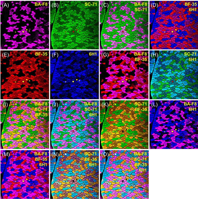
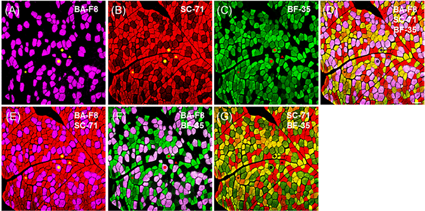
Representative photomicrographs of bovine muscle reacted with four monoclonal antibodies are presented in Fig. 1. Three pure types, I, IIA, and IIX, were identified by a combination of four anti-MHCs, including BA-F8, SC-71, BF-35, and 6H1, as shown in Table 3. Previously, those antibodies had been adopted to identify muscle fiber types regardless of species (Table 2). Muscle fiber type IIB was not detected by any anti-MHC antibodies in the major bovine muscles (LT, PM, SM, and ST), as is consistent with previous studies: MHC 2b isoform (corresponding to type IIB) was not detected at the gene (myosin 4) as well as protein levels in skeletal muscles of adult cows and steers (Chikuni et al., 2004;Kim et al., 2016). Three pure types were easily identified by the combination of two anti-MHC antibodies: BA-F8 and BF-35 (Fig. 1G); SC-71 and 6H1 (Fig. 1H); SC-71 and BF-35 (Fig. 1K); BA-F8 and 6H1 (Fig. 1L). In addition, combinations of three or more anti-MHC antibodies can be used to identify three pure types, as shown in Fig. 1 (I, J, M, N, and O). Hybrid fiber types such as I+IIA and IIA+IIX could be expected from the bovine muscles. Hybrid type I+IIA can be identified by a combination of BA-F8, SC-71, and 6H1 (Fig. 1J); however, this type was not found in the present study (Fig. 1). Another hybrid fiber type, IIA+IIX, was detected by a combination of BF-35 and 6H1 (Fig. 1D). Additionally, the combinations with three antibodies, such as a set of BA-F8, BF-35, and 6H1 (Fig. 1M) or a set of SC-71, BF-35, and 6H1 (Fig. 1N), allowed to detect hybrid fiber type IIA+IIX. Although hybrid fibers were confirmed with those combinations, their fluorescence intensities were lower than that of pure types, as observed by Bloemberg and Quadrilatero (2012). To improve the resolution of the hybrid type, secondary antibodies conjugated with other fluorescent dyes were used and the visualized sections are presented in Fig. 2. A combination of three anti-MHC antibodies such as BA-F8, SC-71, and BF-35 was adopted, and Alexa Fluor 594 and 488 were used for detection of SC-71 and BF-35, respectively. Consequently, the color of hybrid type IIA+IIX can be distinguished from those of the fiber types I, IIA, and IIX (Fig. 2D and 2G).

kosfa-40-1-132-g1
Fig. 1. Result of multicolor immunofluorescence analysis using four anti-myosin heavy chain antibodies (BA-F8, SC-71, BF-35, and 6H1) in bovine longissimus thoracis muscle. Primary and secondary antibodies were applied using cocktail procedures (see Table 1). The images are shown as a single (A, B, E, and F) or merged ones (C, D, G, H, I, J, K, L, M, N, and O), and antibodies specific to myosin heavy chains are presented on each image. Muscle fiber types: •, I; ♦, IIA; ▴, IIX; ⋆, IIAX. Bar=100 μm.
Download Original Figure
Table 3. Reactivity of monoclonal antibodies to myosin heavy chain (MHC) isoforms and identification of muscle fiber types
Antibodies Muscle fiber types
I (MHC slow/I) I+IIA (MHCs I/slow & 2a) IIA (MHC 2a) IIA+IIX (MHCs 2a & 2x) IIX (MHC 2x) IIX+IIB (MHCs 2x & 2b) IIB (MHC 2b)
Bovine skeletal muscles in cocktail
 BA-F8 +++
 SC-71 +++ ++ +(+)
 BF-35 ++ +++ +
 6H1 + ++ +++
Porcine skeletal muscles in cocktail
 BA-F8 +++
 SC-71 ++ +++ + + +
 BF-35 +++ +++ +
 BF-F3 + +++
Porcine skeletal muscles in a series
 BA-F8 +++
 SC-71 +++ ++ + +
 BF-35 ++ +++ +
 BF-F3 + +++

Positive reactivity: +, weak; ++, moderate; +++, strong. Negative reactivity: −.

Download Excel Table
kosfa-40-1-132-g2
Fig. 2. Result of multicolor immunofluorescence analysis using three anti-myosin heavy chain antibodies (BA-F8, SC-71, and BF-35) in bovine longissimus thoracis muscle. Primary and secondary antibodies were applied using cocktail procedures (see Table 1). The images are shown as a single (A, B, and C) or merged ones (D, E, F, and G), and antibodies specific to myosin heavy chains are presented on each image. Muscle fiber types: •, I; ♦, IIA; ▴, IIX; ⋆, IIAX. Bar=100 μm.
Download Original Figure
Porcine muscle fiber type identification

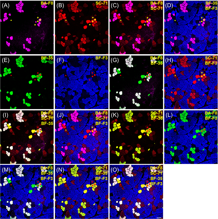
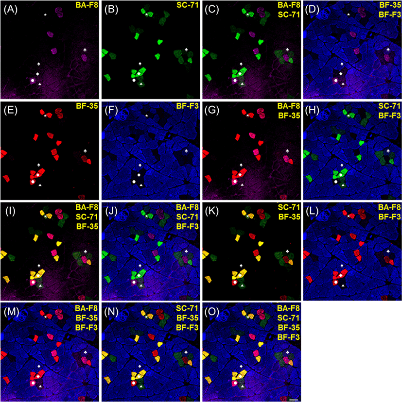
The results of immunofluorescence of porcine muscle, which were reacted by four monoclonal antibodies, such as BA-F8, SC-71, BF-35, and BF-F3, are presented in Figs. 3 and 4. Muscle fiber type IIX in bovine muscle was identified by 6HI antibody; however, this antibody did not work in any porcine muscles. Thus, 6H1 was not adopted for muscle fiber typing of porcine muscles. Among the four anti-MHC antibodies, BA-F8, BF-35, and BF-F3 showed the same specificity to MHCs as previously observed in porcine muscles (Kim et al., 2014; Lefaucheur et al., 2002; Quiroz-Rothe and Rivero, 2004). However, SC-71 had a different reactivity to MHCs different from that in the previous studies. In porcine skeletal muscles, SC-71 generally reacted with MHCs IIA and IIX with different intensities (Kim et al., 2013; Lefaucheur et al., 2002). In the present study, SC-71 reacted with MHC I as well as MHCs IIA and IIX (Fig. 3B). Thus, hybrid fiber type I+IIA could not be identified. From the sections with serial staining procedure, the normal reactivity of SC-71 was observed to have strong intensity with type IIA and weak intensity with type IIX (Fig. 4B). Four pure types (I, IIA, IIX, and IIB) could be detected by combinations of two or more anti-MHC antibodies: BA-F8 and SC-71 (Figs. 3C and 4C); BA-F8, SC-71, and BF-35 (Figs. 3I and 4I); BA-F8, SC-71, and BF-F3 (Figs. 3J and 4J); BA-F8, BF-35, and BF-F3 (Figs. 3M and 4M); BA-F8, SC-71, BF-35, and BF-F3 (Figs. 3O and 4O). The expectable hybrid fiber types were I+IIA, IIA+IIX, and IIX+IIB; however, IIA+IIX and IIX+IIB were detected in the present study. Hybrid type IIA+IIX can be distinguished by a combination of SC-71 and BF-35, whereas type IIX+IIB can be identified with a combination of SC-71 and BF-F3. Among the hybrid fiber types, those two types can be confirmed using both cocktail and serial staining procedures. Thus, our findings indicate that hybrid fiber types IIA+IIX and IIX+IIB can be identified by both cocktail and serial staining procedures, but hybrid fiber type I+IIA can be detected not by cocktail but by serial staining procedure. In other words, the method of cocktail staining could not be recommended for porcine muscle fiber typing despite its time efficiency. To help to distinguish each muscle fiber type, secondary antibodies replaced with other antibodies conjugated with different fluorescent dyes (Alexa Fluor 594 to 488 for SC-71 and 488 to 594 for BF-35) consequently improved resolutions for type I and IIA (Fig. 4O).

kosfa-40-1-132-g3
Fig. 3. Result of multicolor immunofluorescence analysis by a cocktail procedure (see Table 1) using four anti-myosin heavy chain antibodies (BA-F8, SC-71, BF-35, and BF-F3) in porcine longissimus thoracis muscle. The images are shown as a single (A, B, E, and F) or merged ones (C, D, G, H, I, J, K, L, M, N, and O), and antibodies specific to myosin heavy chains are presented on each image. Muscle fiber types: •, I; ♦, IIA; ▴, IIX; ♠, IIB; ♣, IIX+IIB. Bar=100 μm.
Download Original Figure
kosfa-40-1-132-g4
Fig. 4. Result of multicolor immunofluorescence analysis by a serial procedure (see Table 1) using four anti-myosin heavy chain antibodies (BA-F8, SC-71, BF-35, and BF-F3) in porcine longissimus thoracis muscle. Primary and secondary antibodies were applied using cocktail procedures (see Table 1). The images are shown as a single (A, B, E, and F) or merged ones (C, D, G, H, I, J, K, L, M, N, and O), and antibodies specific to myosin heavy chains are presented on each image. Muscle fiber types: •, I; ♦, IIA; ▴, IIX; ♠, IIB; ⋆, IIA+IIX; ♣, IIX+IIB. Bar=100 μm.
Download Original Figure
Muscle fiber characteristics

Muscle fiber size (CSA, μm2) and composition (relative area and relative number, %) was investigated in bovine and porcine skeletal muscles are presented in Table 4. The improved procedures for immunofluorescence by combination of anti-MHC antibodies and fluorescent dyes allowed us to identify four (three pure and one hybrid) fiber types in bovine muscle and six (four pure and two hybrid) fiber types in porcine skeletal muscle. In bovine muscles, hybrid type IIA+IIX was detected with ranges from 1.90% to 4.16% of relative numbers and from 1.97% to 5.16% of relative area. Previously, three fiber types, I, IIA, and IIX (or IIB), were observed in bovine muscles (Hwang et al., 2010; Kim et al., 2016; Ozawa et al., 2000), whereas hybrid fiber types were not frequently identified (Moreno-Sanchez et al., 2008; Greenwood et al., 2009). Fiber type IIB and hybrid type IIX+IIB were not detected in bovine muscle because of the lack of MHC 2b in major bovine muscles (LT, PM, and SM), as previously reported (Kim, 2014; Kim et al., 2016). Muscle fiber composition showed a similar trend to the results of previous reports (Hwang et al., 2010; Ozawa et al., 2000). However, muscle fiber size of LT muscle was not consistent with previous findings. There was no significant (p>0.05) difference in CSA of all fiber types in LT muscle.

Table 4. Muscle fiber characteristics of bovine and porcine skeletal muscles
Muscles Treats Muscle fiber type1) SE
I IIA IIA+IIX IIX IIX+IIB IIB
Bovine skeletal muscles
Psoas major Cross-sectional area (μm2) 2,389B 3,646AB 4,205A 4,806A - - 295.21
Relative area (%) 38.19A 26.94A 5.16B 29.71A - - 4.18
Relative number (%) 51.85A 23.92B 3.96C 20.28B - - 5.74
Longissimus thoracis Cross-sectional area (μm2) 2,706 2,247 2,796 2,756 - - 44.54
Relative area (%) 30.44B 26.57C 3.38D 39.61A - - 3.69
Relative number (%) 29.08B 30.63B 3.12C 37.16A - - 3.72
Semimembranosus Cross-sectional area (μm2) 3,016B 4,095A 4,212A 4,064A - - 219.92
Relative area (%) 26.67B 35.24A 4.74C 33.35A - - 3.27
Relative number (%) 33.11A 32.11A 4.16B 30.61A - - 3.51
Semitendinosus Cross-sectional area (μm2) 3,137C 3,889B 4,365AB 4,724A - - 245.47
Relative area (%) 25.13B 26.05B 1.97C 46.86A - - 4.61
Relative number (%) 31.85B 26.67C 1.90D 39.58A - - 4.07
Porcine skeletal muscles
Psoas major Cross-sectional area (μm2) 2,375C 2,166C - 3,409B - 4,191A 216.04
Relative area (%) 18.47C 20.38BC - 28.76AB - 32.38A 2.18
Relative number (%) 23.48 28.13 - 25.20 - 23.19 1.30
Longissimus thoracis Cross-sectional area (μm2) 2,743C 3,052C 4,765BC 6,869AB 8,251A 6,114AB 438.73
Relative area (%) 6.23C 3.40C 0.44C 17.62B 4.39C 68.14A 4.29
Relative number (%) 12.85B 6.12C 0.57D 14.55B 3.06CD 63.14A 3.97
Semimembranosus Cross-sectional area (μm2) 3,055C 3,011C 6,631A 3,634BC 4,520B 6,997A 359.70
Relative area (%) 6.28B 4.69B 2.91B 7.16B 2.32B 79.26A 5.50
Relative number (%) 11.95B 8.59BC 2.62C 11.35B 2.86C 65.37A 4.17
Semitendinosus Cross-sectional area (μm2) 4,866CD 4,468D 4,992CD 5,407C 7,479A 6,282B 213.76
Relative area (%) 23.89A 14.07B 1.59C 24.78A 5.97BC 29.70A 1.97
Relative number (%) 26.66A 17.03A 1.67B 24.67A 4.32B 25.65A 2.01

1 Muscle fiber types were identified by monoclonal antibodies being specific to myosin heavy chains, as shown in Table 3.

A-D Means±SE with different superscripts are significantly different within a same row (p<0.05).

Download Excel Table

In porcine skeletal muscles, except for PM, hybrid fiber types IIA+IIX and IIX+IIB were identified; they were distributed from 3.63% to 5.99% of relative numbers and from 4.83% to 7.56% of relative area. It has been reported that porcine muscles are composed of 1.7%–11.9% of hybrid fiber types IIA+IIX and IIX+IIB (Kim et al., 2013). The other hybrid type, I+IIA, was previously reported as being observed in rodent, bovine, and canine skeletal muscles (Acevedo and Rivero, 2006; Moreno-Sanchez et al., 2008). However, there was no hybrid type I+IIA in the present study, perhaps because these were different species. Muscle fiber size showed significant differences between type I and type IIB, and type IIB was larger than type I regardless of muscle types (p>0.05). Fiber size of hybrid type IIX+IIB was larger than that of another hybrid type, IIA+IIX, in porcine LT muscle, as is consistent with our previous report (Kim et al., 2014). Unlike LT muscle, SM showed an opposite trend in hybrid fiber size: type IIA+IIX (6,631 μm2 of CSA) was bigger than type IIX+IIB (4,520 μm2 of CSA). The result of muscle fiber compositions (relative area and relative number) of porcine muscles except for ST was consistent with previous studies (Kim et al., 2014; Ryu et al., 2019) showing that relative area or number of type IIB was higher than those of type I, whereas ST was observed to have the same amount of type I as of type IIB. This finding indicates that muscle fiber compositions can be dependent on muscle type.

Conclusion

To optimize typing of muscle fiber in bovine muscle, the combinations of three or four anti-MHC antibodies (BA-F8, SC-71, BF-35, and 6H1) are required, and the cocktail staining procedure with primary and secondary antibodies could save time. Porcine muscle fibers could be typed using four anti-MHC antibodies (BA-F8, SC-71, BF-35, and BF-F3). Unlike for the bovine muscle, cocktail procedure is not recommended in porcine muscle, because of the abnormal reactivity of SC-71. For improved resolution, secondary antibodies conjugated with different fluorescent dyes allowed us to distinguish individual types of muscle fiber in bovine as well as porcine muscles. Accordingly, four muscle fiber types, including one hybrid type, were identified in bovine muscles, and six fiber types (four pure types and two hybrid types) were detected in porcine muscles. However, the hybrid fiber type I+IIA was not found in either species. Differentially distributed muscle fiber types were dependent on species and muscle type.

Notes

Conflict of Interest

The authors declare no potential conflict of interest.

Acknowledgements

This work was supported by Research Resettlement Fund for the new faculty of Seoul National University and by the National Research Foundation of Korea (NRF) grant funded by Korea government (MSIT) (NRF-2019R1C1C1011056).

Notes

Author Contributions

Conceptualization: Kim GD. Data curation: Kim GD, Song SM. Formal analysis: Song SM, Anh CH. Methodology: Song SM, Ahn CH. Writing – original draft: Song SM. Writing – review & editing: Song SM, Ahn CH, Kim GD.

Notes

Ethics Approval

This article does not require IRB/IACUC approval because there are no human and animal participants.

References

1.

Acevedo LM, Rivero J LL. 2006; New insights into skeletal muscle fibre types in the dog with particular focus towards hybrid myosin phenotypes. Cell Tissue Res. 323:283-303

2.

Arguello A, Lopez-Fernandez JL, Rivero JLL. 2001; Limb myosin heavy chain isoproteins and muscle fiber types in the adult goat (Capra hircus). Anat Rec. 264:284-293

3.

Blanco CE, Sieck GC, Edgerton VR. 1988; Quantitative histochemical determination of succinic dehydrogenase activity in skeletal muscle fibres. Histochem J. 20:230-243

4.

Bloemberg D, Quadrilatero J. 2012; Rapid determination of myosin heavy chain expression in rat, mouse, and human skeletal muscle using multicolor immunofluorescence analysis. PLOS ONE. 7:e35273

5.

Brooke MH, Kaiser KK. 1970; Three “myosin adenosine triphosphatase” system: The nature of their pH liability and sulfhydryl dependence. J Histochem Cytochem. 18:670-672

6.

Chang KC, da Costa N, Blackley R, Southwood O, Evans G, Plastow G, Wood JD, Richardson RI. 2003; Relationships of myosin heavy chain fibre types to meat quality traits in traditional and modern pigs. Meat Sci. 64:93-103

7.

Chikuni K, Muroya S, Nakajima I. 2004; Myosin heavy chain isoforms expressed in bovine skeletal muscles. Meat Sci. 67:87-94

8.

Choi YM, Ryu YC, Kim BC. 2007; Influence of myosin heavy- and light chain isoforms on early postmortem glycolytic rate and pork quality. Meat Sci. 76:281-288

9.

Eggert JM, Depreux FF, Schinckel AP, Grant AL, Gerrard DE. 2002; Myosin heavy chain isoforms account for variation in pork quality. Meat Sci. 61:117-126

10.

Gil M, Serra X, Gispert M, Oliver MA, Sanudo C, Panea B, Olleta JL, Campo M, Olivan M, Osoro K, Garcia-Cachan MD, Cruz-Sagredo R, Izquierdo M, Espejo M, Martin M, Piedrafita J. 2001; The effect of breed-production systems on the myosin heavy chain 1, the biochemical characteristics and the colour variables of Longissimus thoracis from seven Spanish beef cattle breeds. Meat Sci. 58:181-188

11.

Gonzalez JM, Dijkhuis RD, Johnson DD, Carter JN, Johnson SE. 2008; Differential response of cull cow muscles to the hypertrophic actions of ractopamine-hydrogen chloride. J Anim Sci. 86:3568-3574

12.

Gorza L. 1990; Identification of a novel type 2 fiber population in mammalian skeletal muscle by combined use of histochemical myosin ATPase and anti-myosin monoclonal antibodies. J Histochem Cytochem. 38:257-265

13.

Graziotti GH, Rios CM, Rivero JL. 2001; Evidence for three fast myosin heavy chain isoforms in type II skeletal muscle fibers in the adult llama (Lama glama). J Histochem Cytochem. 49:1033-1044

14.

Graziotti GH, Palencia P, Delhon G, Rivero JL. 2004; Neuromuscular partitioning, architectural design, and myosin fiber types of the M. vastus lateralis of the llama (Lama glama). J Morphol. 262:667-681

15.

Greenwood PL, Tomkins NW, Hunter RA, Allingham PG, Harden S, Harper GS. 2009; Bovine myofiber characteristics are influenced by postweaning nutrition. J Anim Sci. 87:3114-3123

16.

Guth L, Samaha FJ. 1969; Qualitative differences between actomyosin ATPase of slow and fast mammalian muscle. Exp Neurol. 25:138-152

17.

Hwang YH, Kim GD, Jeong JY, Hur SJ, Joo ST. 2010; The relationship between muscle fiber characteristics and meat quality traits of highly marbled Hanwoo (Korean native cattle) steers. Meat Sci. 86:456-461

18.

Kim GD. 2014; Analysis of myosin heavy chain isoforms from longissimus thoracis muscle of Hanwoo steer by electrophoresis and LC-MS/MS. Korean J Food Sci Anim Resour. 34:656-664

19.

Kim GD, Ryu YC, Jeong JY, Yang HS, Joo ST. 2013; Relationship between pork quality and characteristics of muscle fibers classified by the distribution of myosin heavy chain isoforms. J Anim Sci. 91:5525-5534

20.

Kim GD, Ryu YC, Jo C, Lee JG, Yang HS, Jeong JY, Joo ST. 2014; The characteristics of myosin heavy chain-based fiber types in porcine longissimus dorsi muscle. Meat Sci. 96:712-718

21.

Kim GD, Yang HS, Jeong JY. 2016; Comparison of characteristics of myosin heavy chain-based Fiber and meat quality among four bovine skeletal muscles. Korean J Food Sci Anim Resour. 36:819-828

22.

Larzul C, Lefaucheur L, Ecolan P, Gogue J, Talmant A, Sellier P, Le Roy P, Monin G. 1997; Phenotypic and genetic parameters for longissimus muscle fiber characteristics in relation to growth, carcass, and meat quality traits in large white pigs. J Anim Sci. 75:3126-3137

23.

Lefaucheur L, Ecolan P, Plantard L, Gueguen N. 2002; New insights into muscle fiber types in the pig. J Histochem Cytochem. 50:719-730

24.

Lucas CA, Kang LH, Hoh JF. 2000; Monospecific antibodies against the three mammalian fast limb myosin heavy chains. Biochem Biophys Res Commun. 272:303-308

25.

Martin TP, Vailas AC, Durivage JB, Edgerton VR, Castleman KR. 1985; Quantitative histochemical determination of muscle enzymes: Biochemical verification. J Histochem Cytochem. 33:1053-1059

26.

Micol D, Oury MP, Picard B, Hocquette JF, Briand M, Dumont R, Egal D, Jailler R, Dubroeucq H, Agabriel J. 2009; Effect of age at castration on animal performance, muscle characteristics and meat quality traits in 26-month-old Charolais steers. Livestock Sci. 120:116-126

27.

Moreno-Sanchez N, Diaz C, Carabano MJ, Rueda J, Rivero JL. 2008; A comprehensive characterisation of the fibre composition and properties of a limb (Flexor digitorum superficialis, membri thoraci) and a trunk (Psoas major) muscle in cattle. BMC Cell Biol. 9:67-81

28.

Ozawa S, Mitsuhashi T, Mitsumoto M, Matsumoto S, Itoh N, Itagaki K, Kohno Y, Dohgo T. 2000; The characteristics of muscle fiber types of longissimus thoracis muscle and their influences on the quantity and quality of meat from Japanese Black steers. Meat Sci. 54:65-70

29.

Pette D, Staron RS. 2000; Myosin isoforms, muscle fiber types, and transitions. Microsc Res Technol. 50:500-509

30.

Quiroz-Rothe E, Rivero JL. 2004; Coordinated expression of myosin heavy chains, metabolic enzymes, and morphological features of porcine skeletal muscle fiber types. Microsc Res Technol. 65:43-61

31.

Rivero JL, Talmadge RJ, Edgerton VR. 1996; Correlation between myofibrillar ATPase activity and myosin heavy chain composition in equine skeletal muscle and the influence of training. Anat Rec. 246:195-207

32.

Ryu YC, Choi YM, Lee SH, Shin HG, Choe JH, Kim JM, Hong KC, Kim BC. 2008; Comparing the histochemical characteristics and meat quality traits of different pig breeds. Meat Sci. 80:363-369

33.

Ryu YC, Lee EA, Chai HH, Park JE, Kim JM. 2019; Effects of a novel p.A41P mutation in the swine myogenic factor 5 (MYF5) gene on protein stabilizing, muscle fiber characteristics and meat quality. Food Sci Anim Resour. 38:711-717.

34.

Schiaffino S, Gorza L, Sartore S, Saggin L, Ausoni S, Vianello M, Gundersen K, Lomo T. 1989; Three myosin heavy chain isoforms in type 2 skeletal muscle fibres. J Muscle Res Cell Motil. 10:197-205

35.

Serrano AL, Perez M, Luca A, Chicharro JL, Quiroz-Rothe E, Rivero JL. 2001; Immunolabelling, histochemistry and in situ hybridisation in human skeletal muscle fibres to detect myosin heavy chain expression at the protein and mRNA level. J Anat. 199:329-337

36.

Talmadge RJ, Grossman EJ, Roy RR. 1996; Myosin heavy chain composition of adult feline (Felis catus) limb and diaphragm muscles. J Exp Zool. 275:413-420

37.

Wojtysiak D, Poltowicz K. 2014; Carcass quality, physico-chemical parameters, muscle fibre traits and myosin heavy chain composition of M. longissimus lumborum from Pu?awska and Polish Large White pigs. Meat Sci. 97:395-403