ARTICLE

Duck Oil-loaded Nanoemulsion Inhibits Senescence of Angiotensin II-treated Vascular Smooth Muscle Cells by Upregulating SIRT1

Eun Sil Kang1https://orcid.org/0000-0003-2946-8400, Hyo Juong Kim2https://orcid.org/0000-0003-2819-0822, Sung Gu Han1https://orcid.org/0000-0002-1485-861X, Han Geuk Seo1,*https://orcid.org/0000-0002-9123-3816
Author Information & Copyright
1Department of Food Science and Biotechnology of Animal Resources, College of Sang-Huh Life Science, Konkuk University, Seoul 05029, Korea
2Taekyung Food and Processing R&D Center, Seoul 07057, Korea
*Corresponding author: Han Geuk Seo, Department of Food Science and Biotechnology of Animal Resources, College of Sang-Huh Life Sciences, Konkuk University, Seoul 05029, Korea, Tel: +82-2-450-0428, Fax: +82-2-455-1044, E-mail: hgseo@konkuk.ac.kr

© Korean Society for Food Science of Animal Resources. This is an Open-Access article distributed under the terms of the Creative Commons Attribution Non-Commercial License (http://creativecommons.org/licenses/by-nc/3.0) which permits unrestricted non-commercial use, distribution, and reproduction in any medium, provided the original work is properly cited.

Received: Jul 02, 2019 ; Revised: Oct 14, 2019 ; Accepted: Dec 02, 2019

Published Online: Jan 01, 2020

Abstract

Cellular senescence is associated with age-related vascular disorders and has been implicated in vascular dysfunctions. Here, we show that duck oil-loaded nanoemulsion (DO-NE) attenuates premature senescence of vascular smooth muscle cells (VSMCs) triggered by angiotensin II (Ang II). Compared with control nanoemulsion (NE), DO-NE significantly inhibited the activity of senescence-associated β-galactosidase, which is a biomarker of cellular senescence, in Ang II-treated VSMCs. SIRT1 protein expression was dose- and time-dependently induced in VSMCs exposed to DO-NE, but not in those exposed to NE, and SIRT1 promoter activity was also elevated. Consistently, DO-NE also dose-dependently rescued Ang II-induced repression of SIRT1 expression, indicating that SIRT1 is linked to the anti-senescence action of DO-NE in VSMCs treated with Ang II. Furthermore, the SIRT1 agonist resveratrol potentiated the effects of DO-NE on VSMCs exposed to Ang II, whereas the SIRT1 inhibitor sirtinol elicited the opposite effect. These findings indicate that DO-NE inhibits senescence by upregulating SIRT1 and thereby impedes vascular aging triggered by Ang II.

Keywords: angiotensin II; duck oil; nanoemulsion; senescence; SIRT1

Introduction

Cellular senescence, a permanent and irreversible state of cell cycle arrest, show distinctive phenotypic changes in morphology and gene expression (Hayflick, 1965; Pazolli and Stewart, 2008). Following a limited number of cell divisions, primary cells undergo replicative senescence characterized by accelerated attrition of telomeres that eventually lead to the incomplete chromosomal replication (Harley et al., 1990). Unlike replicative senescence, stress-induced premature senescence is induced by diverse factors that cause cellular stress such as angiotensin II (Ang II), ultraviolet radiation, and hydrogen peroxide (Toussaint et al., 2000; Touyz and Schiffrin, 2000). Recently, Ang II was reported to trigger aging of vascular smooth muscle cells (VSMCs) by causing oxidative DNA damage which is intimately linked to the stability of atherosclerotic plaques (Herbert et al., 2008; Matthews et al., 2006). These findings are consistent with the reports that blockade of Ang II activity by polyphenols, such as resveratrol and those found in berries, inhibits vascular senescence-mediated intracellular signaling, leading to blockade of vascular age-associated diseases including atherosclerosis (Feresin et al., 2016; Kim et al., 2018; Najjar et al., 2005). Thus, aging-related vascular disorders may be prevented by controlling cellular senescence. As a potential candidate among diverse anti-senescence factors, the NAD-dependent deacetylase SIRT1 has a pivotal function in cardiovascular systems and is highly expressed (Potente et al., 2007). SIRT1 elicits beneficial effects on neointima formation, vascular remodeling, and atherosclerosis by inhibiting stress-induced cellular senescence (Gao et al., 2014; Kim et al., 2012; Li et al., 2011b). Exacerbated DNA damage and senescence are observed in VSMCs located in atherosclerotic regions, in which SIRT1 expression is reduced (Gorenne et al., 2013, Zhang et al., 2008). In addition, our previous studies showed that peroxisome proliferator-activated receptor δ-mediated induction of SIRT1 expression suppresses Ang II-triggered premature senescence of human VSMCs and endothelial cells (Kim et al., 2011; Kim et al., 2012). Thus, molecules that upregulate expression of the anti-senescence protein SIRT1 alter the pathological cardiovascular conditions caused by aging of vascular cells (Gorenne et al., 2013; Ota et al., 2008).

Duck oil is an avian oil that derived from duck skin, a by-product of duck meat processes (Shin et al., 2019). Recent report has shown that duck skin-derived oil contains a higher amount of long-chain fatty acids including oleic acid (18:1) and linoleic acid (18:2) than other animal skin fats, such as chicken, swine, bovine (Shin et al., 2019). In fact, long-chain fatty acids have been shown direct beneficial effects in the prevention and treatment of many diseases, such as diabetes, obesity, and cardiovascular disorders (Fuke and Nornberg, 2017; Massaro and De Caterina, 2002). Furthermore, duck oil showed a high unsaturated fatty acid/saturated fatty acid ratio (above 50%) compared with fats derived from swine and bovine, indicating usefulness of duck oil in food industries (Shin et al., 2019). However, the biological activity of duck oil has not been experimentally elucidated. Consequently, we investigated the effects of duck oil in the vascular aging processes. We here demonstrate that duck oil derived from duck skin inhibits premature senescence of VSMCs triggered by Ang II by upregulating SIRT1.

Materials and Methods

Oil extraction from duck skin

Duck skin was obtained from Farm Duck Co. (Jeongeup-si, Korea). A pressurized hot water extraction method was used to isolate the oil as described previously (Plaza & Turner, 2015). In detail, skin samples from eight individual duck were washed several times with distill water and connective tissues and visible fat were removed from the skin. Following these procedures, a pressure extractor was applied to extract oil from the skin by heating the skin at 115°C under 1.4 kgf/cm2. Following treatment for 3 h, the samples were dehydrated for 30 min and then filtrated with a 40-mesh bucket filter. Finally, the oil phase (supernatant) was obtained by centrifugation of the filtrate at 14,500×g for 1 h.

Preparation of nanoemulsions

Duck oil-loaded nanoemulsion (DO-NE) was generated by mixing 20% (w/w) duck oil in medium chain triglyceride oil (NOW Foods, Elmhurst, IL, USA) containing the hydrophobic emulsifier soy lecithin (ESFOOD, Gunpo, Korea). Distilled water was mixed with the hydrophilic emulsifier Tween-80 (Daejung Chemical, Seoul, Korea) to generate the aqueous phase. After mixing using a magnetic bar for 2 h, the samples were sequentially homogenized (550×g, 10 min), ultrasonicated (15 min), and exposured to high pressure (10,000 psi, three times) to prepare nanoemulsions (NE). The same procedure without duck oil was performed to generate control NE.

Characterization of nanoemulsions

A particle size and zeta potential analyzer was used to determine the polydispersity index (PDI), mean droplet size, and zeta potential of NEs. Specifically, a polystyrene latex cell containing 1 mL NE was analyzed using a detector angle of 90° and a wavelength of 633 nm at 25°C to determine PDI and mean droplet size. A droplet of diluted NE was placed on a copper grid and briefly dried. After negative staining with phosphotungstic acid, the grid was dried overnight and then images were acquired using a transmission electron microscope at 110 kV and at a magnification of 70 K.

Cell culture

Primary VSMCs were isolated from the thoracic aorta of a rat as described previously (Hwang et al., 2016). Briefly, the aorta of a male Sprague-Dawley rat was dissected longitudinally. Endothelial cells were removed and the medial layer of the aorta was chopped into small fragments and incubated up to 3 d. Fresh medium was then added, and fragmented tissues were incubated for several further days. Thereafter, migrated VSMCs were collected and passaged in Dulbecco’s modified Eagle’s medium containing antibiotics and 10% fetal bovine serum using routine cell culture procedures. VSMCs employed in this study were originally isolated in 2008. Cells at passage 4-8 were used in the present study.

Cytotoxicity assay

The MTT assay was performed to assess cell viability as described previously (Yoo et al., 2016). Specifically, the indicated concentration of NE or DO-NE was added to VSMCs plated into 24-well plates, and cultures were incubated for 24 h. Thereafter, the cells were further incubated in culture medium containing 0.1 mg/mL MTT for 2 h. After removal of the medium, crystalized formazan was dissolved in acidified isopropanol and the optical density was measured at 570 nm.

SA β-gal staining

VSMCs cultured in 60 mm dishes were pretreated with NE or DO-NE for 24 h. Thereafter, the cells were exposed to vehicle (Dimethyl sulfoxide, DMSO) or Ang II for 72 h, washed briefly with cold phosphate-buffered saline, and then stained with a solution for 24 h at 37°C as described previously (Kim et al., 2012). Fluorescence microscopy was performed to detect stained cells. The number of stained cells in four individual captured images was counted on a computer monitor using a DS-Ri2 camera equipped to Nikon Eclipse Ti2 inverted fluorescence microscope (Tokyo, Japan) by an independent observer.

Western blot analysis

SDS-polyacrylamide gel electrophoresis was performed using whole-cell lysates of VSMCs treated with the indicated reagents, and then immunoblotting was conducted using specific antibodies. Specifically, the membranes were blocked with 3% skim milk prepared in Tris-buffered saline containing 0.1% Tween-20 for 2 h at ambient temperature. Thereafter, the membranes were reacted with a specific antibody at 4°C overnight and then with a secondary antibody conjugated with peroxidase at room temperature for 2 h. Signals were detected by enhanced chemiluminescence as described previously (Hwang et al., 2016). The band intensity of blots was quantified by image J software (NIH, Bethesda, MD, USA) and normalized using loading control.

Reporter gene assay

The mouse-specific SIRT1 promoter (−2,487 to −30 in pGL4) was obtained from Dr. Toren Finkel [National Institutes of Health (NIH), MD, USA]. VSMCs were co-transfected with the SIRT1 luciferase reporter plasmid (1 μg) and the SV40 β-galactosidase expression vector (0.5 μg) and incubated for 24 h. Following exposure to different concentrations of DO-NE for 72 h, the cells were lysed and promoter activity was measured. β-galactosidase activity was used to normalize variations in the transfection efficiency between samples.

Statistical analysis

Statistical significance (n=3 or 4) was determined by performing a one-way ANOVA with the Bonferroni post-hoc test using SigmaPlot 12.0.

Results and Discussion

Physicochemical properties of nanoemulsions

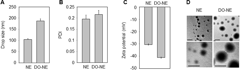
The solubility and biological performance of nanoparticles are affected by the physicochemical properties of NEs (Ghasemiyeh and Mohammadi-Samani, 2018). Therefore, we analyzed the zeta potential, PDI, and droplet size of the NEs. The mean droplet size and zeta potential of the NEs ranged from 104 to 192 nm and from −30 to −41 mV, respectively, and the NEs were of a uniform size and had a spherical shape without any flocculation (Fig. 1).

kosfa-40-1-106-g1
Fig. 1. Properties of NE and DO-NE. (A–C) Mean droplet size (A), PDI (B), and zeta potential (C) of NE or DO-NE loaded on a polystyrene latex cell were measured at 25°C with a detector angle of 90° at 633 nm using a zeta potential and particle size analyzer. Each sample was measured at least three times and the average values were calculated. (D) Transmission electron microscopy images. Bars, 500 nm. DO-NE, duck oil-loaded nanoemulsion; PDI, polydispersity index.
Download Original Figure
Effect of DO-NE on Ang II-triggered senescence of VSMCs

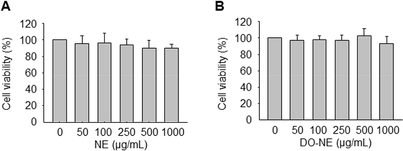
To determine the optimal concentrations of the NEs for treatment of VSMCs, the MTT assay was performed to assess cell viability. Treatment with increasing concentrations (0, 50, 100, 250, 500, and 1,000 μg/mL) of NE and DO-NE for 24 h did not significantly affect cell viability (Fig. 2). Accordingly, the NEs were used at a concentration of 1 mg/mL in subsequent experiments.

kosfa-40-1-106-g2
Fig. 2. Effects of NE and DO-NE on viability of VSMCs. (A–B) Cells were exposed to different concentrations (0, 50, 100, 250, 500, or 1,000 μg/mL) of NE (A) or DO-NE (B) for 24 h. Cell viability was measured using the MTT assay. Results are expressed as means±SE (n=4). DO-NE, duck oil-loaded nanoemulsion; VSMCs, vascular smooth muscle cells.
Download Original Figure

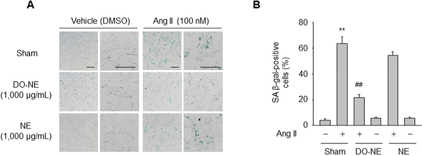
Next, we examined whether DO-NE affects Ang II-triggered senescence of VSMCs. SA β-gal activity, a senescence biomarker, in the sham group was significantly higher in VSMCs treated with Ang II (63.63±5.17, p<0.01) for 3 days than in cells exposed to DMSO (4.22±1.09). However, this increase was significantly suppressed by treatment with DO-NE (21.77±2.38, p<0.01) but not by treatment with NE (54.42±2.76; Fig. 3A–B). These findings are consistent with the previous report that oils from avian species, such as emu and duck, contain a high ratio of unsaturated fatty acids to saturated fatty acids and protect against oxidative damage in a model biological membrane system (Bennett et al., 2008). Additionally, the triglyceride concentration in the serum and liver of rats is decreased more by feeding of duck oil than by feeding of soybean oil (Koh et al., 1995). Although the molecular basis of the anti-senescence effects of duck oil in the vascular system is unknown, our results suggest that duck oil elicits beneficial effects on senescence of VSMCs stimulated by Ang II and can therefore be potentially used as a functional food.

kosfa-40-1-106-g3
Fig. 3. Effects of NE and DO-NE on Ang II-induced senescence of VSMCs. (A–B) Cells pretreated in the presence or absence of NE or DO-NE for 24 h were exposed to vehicle (DMSO) or Ang II for 72 h. SA β-gal staining was performed to detect senescent cells (A) and quantified. The percentage of SA β-gal-positive cells is plotted (B). Bars, 200 μm. Representative images are shown and data are expressed as means±SE (n=4). ** p<0.01 vs. untreated sham group; ## p<0.01 vs. Ang II-treated sham group. DO-NE, duck oil-loaded nanoemulsion; VSMCs, vascular smooth muscle cells; DMSO, dimethyl sulfoxide.
Download Original Figure
Effect of DO-NE on SIRT1 expression

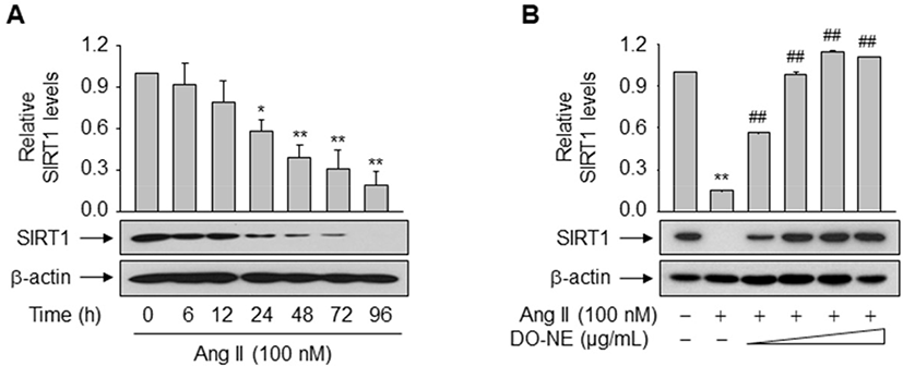
The pro-longevity gene SIRT1 is implicated in lifespan modulation in various organisms ranging from yeast to mammals (Haigis and Sinclair, 2010). SIRT1 was directly implicated in the modulation of cellular aging by deacetylating critical transcription factors such as forkhead box protein and p53, thereby repressing the transcription of Ang II type I receptor as demonstrated in VSMCs (Brunet et al., 2004; Langley et al., 2002; Miyazaki et al., 2008). We examined the effects of duck oil on SIRT1 expression in VSMCs. DO-NE, but not NE, dose- and time-dependently enhanced the expression level of SIRT1 protein, as expected (Fig. 4A–C). The level of SIRT1 protein was highest upon treatment with 1,000 μg/mL DO-NE for 72 h (2.03±0.14, p<0.01). When VSMCs were treated with 1,000 μg/mL DO-NE, SIRT1 expression was significantly increased at 48 h (1.85±0.20, p<0.05) and this persisted for up to 72 h (2.36±0.29, p<0.01).

kosfa-40-1-106-g4
Fig. 4. Effects of NE and DO-NE on SIRT1 expression. (A–C) VSMCs were incubated with the indicated concentrations of NE (A) or DO-NE (B) for 72 h (A) or with 1,000 μg/mL DO-NE for the indicated durations (C). Aliquots of whole-cell lysates were analyzed by western blotting. Band intensities were quantified with an image analyzer. The ratio of SIRT1 to β-actin is plotted (upper panels of B and C). (D) VSMCs co-transfected with 0.5 μg pSV β-Gal and 1 μg of the SIRT1 luciferase reporter plasmid were stimulated with the indicated concentrations of DO-NE for 72 h. Luciferase activity is normalized by β-galactosidase activity. Data are expressed as means±SE (n=3 or 4). * p<0.05, ** p<0.01 vs. untreated group. DO-NE, duck oil-loaded nanoemulsion; VSMCs, vascular smooth muscle cells.
Download Original Figure

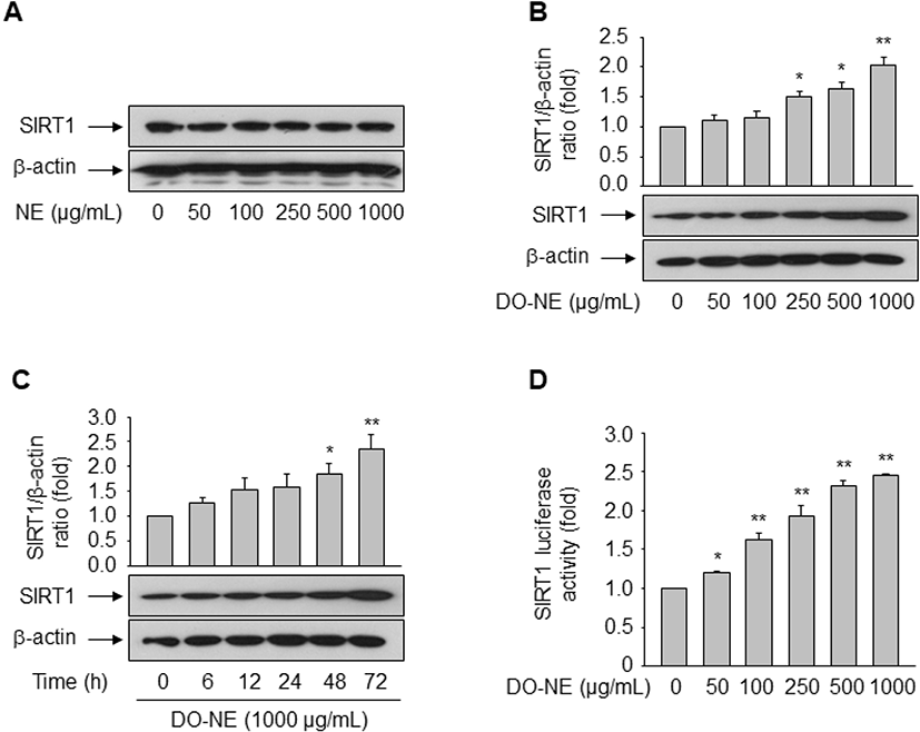
No previous report has shown that duck oil affects expression of SIRT1. Hence, we investigated whether duck oil regulates transcription of the SIRT1 gene. To this end, VSMCs were transfected with a luciferase reporter construct containing the SIRT1 promoter. As shown in Fig. 4D, DO-NE dose-dependently enhanced SIRT1 promoter activity from 50 μg/mL DO-NE (1.21±0.01, p<0.05) to 1,000 μg/mL DO-NE (2.45±0.01, p<0.01). This result is consistent with the induction of SIRT1 protein expression by DO-NE, indicating that duck oil transcriptionally regulates expression of SIRT1 in VSMCs. In fact, SIRT1 is reported to modulate diverse biological processes, including energy metabolism, stress responses, inflammation, and aging (Brunet et al., 2004; Cohen et al., 2004; Feige and Auwerx, 2008; Yeung et al., 2004). In addition to the role of caloric restriction in SIRT1 regulation (Cohen et al., 2004), diverse cellular molecules such as p53, the HIC:CtBP co-repressor complex, E2F1, cAMP response element-binding protein, breast cancer 1, and TLX are associated with modulation of SIRT1 transcription (Chen et al., 2005; Iwahara et al., 2009; Noriega et al., 2011; Wang et al., 2006; Wang et al., 2008; Yi and Luo, 2010). We previously demonstrated that peroxisome proliferator-activated receptor δ, a ligand-dependent nuclear receptor, induces SIRT1 transcription in rat VSMCs and human endothelial cells, thereby regulating cellular senescence and proliferation triggered by the atherogenic-molecule oxidized low-density lipoprotein and Ang II, respectively (Hwang et al., 2016; Kim et al., 2012). Additionally, the present study clearly demonstrates that duck oil also induces SIRT1 expression.

Role of SIRT1 in DO-NE-mediated inhibition of VSMC senescence induced by Ang II

To examine the direct impact of duck oil-mediated induction of SIRT1 on suppression of VSMC senescence triggered by Ang II, expression of endogenous SIRT1 was monitored in VSMCs exposed to Ang II with or without DO-NE. Treatment with Ang II time-dependently suppressed the level of endogenous SIRT1 protein from 24 h (0.58±0.08, p<0.05; Fig. 5A). However, DO-NE dose-dependently reversed this effect from 100 μg/mL DO-NE (p<0.01), indicating that induction of SIRT1 protein is essential for DO-NE-mediated suppression of VSMC senescence triggered by Ang II (Fig. 5B).

kosfa-40-1-106-g5
Fig. 5. Effect of DO-NE on the endogenous SIRT1 level. (A) VSMCs were exposed to Ang II for the indicated durations. (B) VSMCs pretreated with increasing concentrations (0, 100, 250, 500, or 1,000 μg/mL) of DO-NE for 24 h were exposed to Ang II for 72 h. Aliquots of whole-cell lysates were analyzed by western blotting with specific antibodies. Band intensities were quantified with an image analyzer. The ratio of SIRT1 to β-actin is plotted and expressed as means±SE (n=3). * p<0.05, ** p<0.01 vs. untreated group; ## p<0.01 vs. Ang II-treated group. DO-NE, duck oil-loaded nanoemulsion; VSMCs, vascular smooth muscle cells.
Download Original Figure

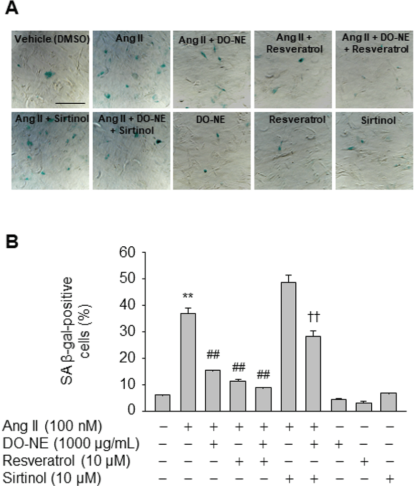
To further elucidate the importance of SIRT1 induction for inhibition of Ang II-triggered VSMC senescence by DO-NE, cells were treated with reagents that modulate SIRT1 activity. The number of SA β-gal-positive cells was lower among VSMCs exposed to Ang II and resveratrol (11.50±0.76, p<0.01) than among VSMCs treated with Ang II alone (37.10±1.95; Fig. 6). Furthermore, the inhibitory effect of DO-NE was potentiated by addition of resveratrol to DO-NE-treated VSMCs (9.03±0.09, p<0.01). By contrast, addition of sirtinol, a specific inhibitor of SIRT1, significantly counteracted the DO-NE-mediated reduction of SA β-gal-positive cells in Ang II-treated cells (28.46±1.89, p<0.01). These observations indicate that the anti-senescence activity of DO-NE in Ang II-treated VSMCs is attributable, at least in part, to DO-NE-mediated induction of SIRT1, a protein that is associated with increased lifespan in mammals (Haigis and Sinclair, 2010). Although the detailed mechanisms by which DO-NE induces expression of SIRT1 are unclear, chemical-mediated modulation of SIRT1 activity clearly indicated that DO-NE inhibits senescence of VSMCs triggered by Ang II in a SIRT1-dependent manner. Consistent with this anti-senescence effect of DO-NE, a previous study demonstrated that expression of a VSMC-specific SIRT1 transgene inhibits Ang II-induced vascular remodeling (Gao et al., 2014). Additionally, ectopic expression of SIRT1 using an adenovirus markedly suppresses hypertrophy of VSMCs triggered by Ang II (Li et al., 2011a). Furthermore, knockout of SIRT1 in VSMCs promotes Ang II-induced vascular cell senescence, thereby accelerating formation and rupture of abdominal aortic aneurysms in apolipoprotein E knockout mice (Chen et al., 2016). These findings suggest that DO-NE-mediated upregulation of SIRT1 is associated with the usefulness of duck oil as a potential functional food to prevent aging-related vascular diseases.

kosfa-40-1-106-g6
Fig. 6. Effects of DO-NE, resveratrol, and sirtinol on senescence of VSMCs triggered by Ang II. (A–B) VSMCs were pretreated with resveratrol or sirtinol for 60 min, exposed to DO-NE for 24 h, and then treated with DMSO or Ang II for 72 h. SA β-gal staining was performed to detect senescent cells (A). Bar, 50 μm. The percentage of SA β-gal-positive cells is plotted (B) and expressed as means±SE (n=4). ** p<0.01 vs. DMSO-treated group; ## p<0.01 vs. Ang II-treated group; †† p<0.01 vs. Ang II + DO-NE-treated group. DO-NE, duck oil-loaded nanoemulsion; VSMCs, vascular smooth muscle cells; DMSO, dimethyl sulfoxide.
Download Original Figure

Conclusion

To obtain scientific evidence of the biological activity of duck oil, we examined the effects of this oil on vascular senescence triggered by Ang II, a vasoactive hormone. The present study demonstrates that DO-NE suppresses Ang II-stimulated senescence of VSMCs by increasing transcription of SIRT1. The present work provides the first clue that induction of SIRT1 by DO-NE suppresses vascular aging. This novel evidence provides insights into the anti-senescence effect of duck oil and the detailed mechanisms underlying SIRT1 expression. The anti-aging activity of duck oil in vascular systems supports the potential use of this oil as a functional food to improve vascular functions.

Notes

Conflict of Interest

The authors declare no potential conflict of interest.

Acknowledgments

This work was supported by Korea Institute of Planning and Evaluation for Technology in Food, Agriculture, Forestry and Fisheries (IPET) through High Value-added Food Technology Development Program, funded by Ministry of Agriculture, Food and Rural Affairs (MAFRA) (2018-118011).

Notes

Author Contributions

Conceptualization: Kang ES, Seo HG. Data curation: Kang ES. Formal analysis: Kang ES, Kim HJ. Methodology: Kang ES, Kim HJ, Seo HG. Software: Kang ES. Validation: Han SG, Seo HG. Investigation: Seo HG. Writing - original draft: Kang ES. Writing - review & editing: Kang ES, Kim HJ, Han SG, Seo HG.

Notes

Ethics Approval

This article does not require IRB/IACUC approval because there are no human and animal participants.

References

1.

Bennett DC, Code WE, Godin DV, Cheng KM. 2008; Comparison of the antioxidant properties of emu oil with other avian oils. Aust J Exp Agric. 48:1345-1350

2.

Brunet A, Sweeney LB, Sturgill JF, Chua KF, Greer PL, Lin Y, Tran H, Ross SE, Mostoslavsky R, Cohen HY, Hu LS, Cheng HL, Jedrychowski MP, Gygi SP, Sinclair DA, Alt FW, Greenberg ME. 2004; Stress-dependent regulation of FOXO transcription factors by the SIRT1 deacetylase. Science. 303:2011-2015

3.

Chen HZ, Wang F, Gao P, Pei JF, Liu Y, Xu TT, Tang X, Fu WY, Lu J, Yan YF, Wang XM, Han L, Zhang ZQ, Zhang R, Zou MH, Liu DP. 2016; Age-associated sirtuin 1 reduction in vascular smooth muscle links vascular senescence and inflammation to abdominal aortic aneurysm. Circ Res. 119:1076-1088

4.

Chen WY, Wang DH, Yen RC, Luo J, Gu W, Baylin SB. 2005; Tumor suppressor HIC1 directly regulates SIRT1 to modulate p53-dependent DNA-damage responses. Cell. 123:437-448

5.

Cohen HY, Miller C, Bitterman KJ, Wall NR, Hekking B, Kessler B, Howitz KT, Gorospe M, de Cabo R, Sinclair DA. 2004; Calorie restriction promotes mammalian cell survival by inducing the SIRT1 deacetylase. Science. 305:390-392

6.

Feige JN, Auwerx J. 2008; Transcriptional targets of sirtuins in the coordination of mammalian physiology. Curr Opin Cell Biol. 20:303-309

7.

Feresin RG, Huang J, Klarich DS, Zhao Y, Pourafshar S, Arjmandi BH, Salazar G. 2016; Blackberry, raspberry and black raspberry polyphenol extracts attenuate angiotensin II-induced senescence in vascular smooth muscle cells. Food Funct. 7:4175-4187

8.

Fuke G, Nornberg JL. 2017; Systematic evaluation on the effectiveness of conjugated linoleic acid in human health. Crit Rev Food Sci Nutr. 57:1-7

9.

Gao P, Xu TT, Lu J, Li L, Xu J, Hao DL, Chen HZ, Liu DP. 2014; Overexpression of SIRT1 in vascular smooth muscle cells attenuates angiotensin II-induced vascular remodeling and hypertension in mice. J Mol Med. 92:347-357

10.

Ghasemiyeh P, Mohammadi-Samani S. 2018; Solid lipid nanoparticles and nanostructured lipid carriers as novel drug delivery systems: Applications, advantages and disadvantages. Res Pharm Sci. 13:288-303

11.

Gorenne I, Kumar S, Gray K, Figg N, Yu H, Mercer J, Bennett M. 2013; Vascular smooth muscle cell sirtuin 1 protects against DNA damage and inhibits atherosclerosis. Circulation. 127:386-396

12.

Haigis MC, Sinclair DA. 2010; Mammalian sirtuins: Biological insights and disease relevance. Annu Rev Pathol. 5:253-295

13.

Harley CB, Futcher AB, Greider CW. 1990; Telomeres shorten during ageing of human fibroblasts. Nature. 345:458-460

14.

Hayflick L. 1965; The limited in vitro lifetime of human diploid cell strains. Exp Cell Res. 37:614-636

15.

Herbert KE, Mistry Y, Hastings R, Poolman T, Niklason L, Williams B. 2008; Angiotensin II-mediated oxidative DNA damage accelerates cellular senescence in cultured human vascular smooth muscle cells via telomere-dependent and independent pathways. Circ Res. 102:201-208

16.

Hwang JS, Ham SA, Yoo T, Lee WJ, Paek KS, Lee CH, Seo HG. 2016; Sirtuin 1 mediates the actions of peroxisome proliferator-activated receptor δ on the oxidized low-density lipoprotein-triggered migration and proliferation of vascular smooth muscle cells. Mol Pharmacol. 90:522-529

17.

Iwahara N, Hisahara S, Hayashi T, Horio Y. 2009; Transcriptional activation of NAD+-dependent protein deacetylase SIRT1 by nuclear receptor TLX. Biochem Biophys Res Commun. 386:671-675

18.

Kim EN, Kim MY, Lim JH, Kim Y, Shin SJ, Park CW, Kim YS, Chang YS, Yoon HE, Choi BS. 2018; The protective effect of resveratrol on vascular aging by modulation of the renin-angiotensin system. Atherosclerosis. 270:123-131

19.

Kim HJ, Ham SA, Kim MY, Hwang JS, Lee H, Kang ES, Yoo T, Woo IS, Yabe-Nishimura C, Paek KS, Kim JH, Seo HG. 2011; PPARδ coordinates angiotensin II-induced senescence in vascular smooth muscle cells through PTEN-mediated inhibition of superoxide generation. J Biol Chem. 286:44585-44593

20.

Kim MY, Kang ES, Ham SA, Hwang JS, Yoo TS, Lee H, Paek KS, Park C, Lee HT, Kim JH, Han CW, Seo HG. 2012; The PPARδ-mediated inhibition of angiotensin II-induced premature senescence in human endothelial cells is SIRT1-dependent. Biochem Pharmacol. 84:1627-1634

21.

Koh JB, Jung BM, Kim JY, Rho MH. 1995; Effects of duck oil on serum and organ lipid composition in mature rats. J Korean Soc Food Nutr. 24:874-879.

22.

Langley E, Pearson M, Faretta M, Bauer UM, Frye RA, Minucci S, Pelicci PG, Kouzarides T. 2002; Human SIR2 deacetylates p53 and antagonizes PML/p53-induced cellular senescence. EMBO J. 21:2383-2396

23.

Li L, Gao P, Zhang H, Chen H, Zheng W, Lv X, Xu T, Wei Y, Liu D, Liang C. 2011a; SIRT1 inhibits angiotensin II-induced vascular smooth muscle cell hypertrophy. Acta Biochim Biophys Sin. 43:103-109

24.

Li L, Zhang HN, Chen HZ, Gao P, Zhu LH, Li HL, Lv X, Zhang QJ, Zhang R, Wang Z, She ZG, Zhang R, Wei YS, Du GH, Liu DP, Liang CC. 2011b; SIRT1 acts as a modulator of neointima formation following vascular injury in mice. Circ Res. 108:1180-1189

25.

Massaro M, De Caterina R. 2002; Vasculoprotective effects of oleic acid: Epidemiological background and direct vascular antiatherogenic properties. Nutr Metab Cardiovasc Dis. 12:42-51

26.

Matthews C, Gorenne I, Scott S, Figg N, Kirkpatrick P, Ritchie A, Goddard M, Bennett M. 2006; Vascular smooth muscle cells undergo telomere-based senescence in human atherosclerosis: Effects of telomerase and oxidative stress. Circ Res. 99:156-164

27.

Miyazaki R, Ichiki T, Hashimoto T, Inanaga K, Imayama I, Sadoshima J, Sunagawa K. 2008; SIRT1, a longevity gene, downregulates angiotensin II type 1 receptor expression in vascular smooth muscle cells. Arterioscler Thromb Vasc Biol. 28:1263-1269

28.

Najjar SS, Scuteri A, Lakatta EG. 2005; Arterial aging: Is it an immutable cardiovascular risk factor?. Hypertension. 46:454-462

29.

Noriega LG, Feige JN, Canto C, Yamamoto H, Yu J, Herman MA, Mataki C, Kahn BB, Auwerx J. 2011; CREB and ChREBP oppositely regulate SIRT1 expression in response to energy availability. EMBO Rep. 12:1069-1076

30.

Ota H, Eto M, Kano MR, Ogawa S, Iijima K, Akishita M, Ouchi Y. 2008; Cilostazol inhibits oxidative stress-induced premature senescence via upregulation of SIRT1 in human endothelial cells. Arterioscler Thromb Vasc Biol. 28:1634-1639

31.

Pazolli E, Stewart SA. 2008; Senescence: The good the bad and the dysfunctional. Curr Opin Genet Dev. 18:42-47

32.

Plaza M, Turner C. 2015; Pressurized hot water extraction of bioactives. Trends Anal Chem. 71:39-54

33.

Potente M, Ghaeni L, Baldessari D, Mostoslavsky R, Rossig L, Dequiedt F, Haendeler J, Mione M, Dejana E, Alt FW, Zeiher AM, Dimmeler S. 2007; SIRT1 controls endothelial angiogenic functions during vascular growth. Genes Dev. 21:2644-2658

34.

Shin DM, Kim DH, Yune JH, Kwon HC, Kim HJ, Seo HG, Han SG. 2019; Oxidative stability and quality characteristics of duck, chicken, swine and bovine skin fats extracted by pressurized hot water extraction. Food Sci Anim Resour. 39:446-458

35.

Toussaint O, Medrano EE, von Zglinicki T. 2000; Cellular and molecular mechanisms of stress-induced premature senescence (SIPS) of human diploid fibroblasts and melanocytes. Exp Gerontol. 35:927-945

36.

Touyz RM, Schiffrin EL. 2000; Signal transduction mechanisms mediating the physiological and pathophysiological actions of angiotensin II in vascular smooth muscle cells. Pharmacol Rev. 52:639-672.

37.

Wang C, Chen L, Hou X, Li Z, Kabra N, Ma Y, Nemoto S, Finkel T, Gu W, Cress WD, Chen J. 2006; Interactions between E2F1 and SirT1 regulate apoptotic response to DNA damage. Nat Cell Biol. 8:1025-1031

38.

Wang RH, Zheng Y, Kim HS, Xu X, Cao L, Luhasen T, Lee MH, Xiao C, Vassilopoulos A, Chen W, Gardner K, Man YG, Hung MC, Finkel T, Deng CX. 2008; Interplay among BRCA1, SIRT1, and Survivin during BRCA1-associated tumorigenesis. Mol Cell. 32:11-20

39.

Yeung F, Hoberg JE, Ramsey CS, Keller MD, Jones DR, Frye RA, Mayo MW. 2004; Modulation of NF-kappaB-dependent transcription and cell survival by the SIRT1 deacetylase. EMBO J. 23:2369-2380

40.

Yi J, Luo J. 2010; SIRT1 and p53, effect on cancer, senescence and beyond. Biochim Biophys Acta. 1804:1684-1689

41.

Yoo T, Ham SA, Hwang JS, Lee WJ, Paek KS, Oh JW, Kim JH, Do JT, Han CW, Kim JH, Seo HG. 2016; Peroxisome proliferator-activated receptor δ inhibits Porphyromonas gingivalis lipopolysaccharide-induced activation of matrix metalloproteinase-2 by downregulating NADPH oxidase 4 in human gingival fibroblasts. Mol Oral Microbiol. 31:398-409

42.

Zhang QJ, Wang Z, Chen HZ, Zhou S, Zheng W, Liu G, Wei YS, Cai H, Liu DP, Liang CC. 2008; Endothelium-specific overexpression of class III deacetylase SIRT1 decreases atherosclerosis in apolipoprotein E-deficient mice. Cardiovasc Res. 80:191-199