


Introduction
With the westernization of food culture, dietary choices in South Korea have changed dramatically. According to statistics, the grain intake in 1998 was 337.2 g/day and it decreased to 292.5 g/day in 2009, while animal foodstuff such as meat, eggs, shellfish, and milk increased from 238.0 g/day in 1998 to 269.3 g/day in 2009 (Ministry of Food and Drug Safety, 2012). Currently, veterinary drugs are widely used not only for the simple prevention and treatment of diseases, but also to improve livestock productivity. As a result, there is an increasing problem of drug residue in food sources. In order to reduce the risk of residual veterinary drug contamination in food, it is necessary to monitor the residual level of veterinary drugs and setting and operation of the scientific withdrawal period (Shin, 2005). Owing to the long-term misuse of veterinary drugs (e.g., antibiotics) involving livestock animals, residual level of these synthetic compounds can be detected in processed products. Consumers are concerned about the long-term effect of ingesting these compounds, especially the possibility of developing drug resistance after a pro-longed low-dosage exposure; thus potentially negating the therapeutic effects of these drugs in patients. Antibiotic resistance is a problem directly related to national health interest and the quality of life. As such, there must be an emphasis on the systematic management, proper distribution and use, as well as safety management of medicine and medical supplies for animals (Shin and Park, 2006). While residual veterinary drugs in food does not immediately lead to the emergence of drug-resistant bacteria, an increase in the incidence of drug resistance could potentially be directly, or indirectly, related to the uncontrolled use of veterinary drugs (Masakzu et al., 2003). Livestock or farmed fish are exposed to veterinary drugs via absorptions through oral and parenteral paths (e.g., injections, skin, and inhalation and the absorbed substances have high affinity to various organs and tissues in animals (i.e., quick accumulation and long elimination time). Globally, there is an increasing interest in veterinary drug resistance, particularly the propagation of tolerance for meat-derived drugs for animals (Ministry of Food and Drug Safety, 2012). Since Korea began exporting pork products to Japan in 1989, 27 maximum residue limits (MRL) involving veterinary drugs in livestock products, along with 57 test methods, have been set as national standards for drug residue evaluation. As of 2014, 130 Korean dairy and marine products are regulated for their veterinary residual drug content. The Ministry of Food and Drug Safety has carried out studies on the pre-to-post monitoring of veterinary drugs in animals since 2005, upon which residual standards and new test methods have been established. A number of test methods were made available with the rapid establishment of MRLs since 2007. As the worldwide standards for the use of veterinary drugs are tightened according to Codex Standards (affiliated with the World Health Organization and Food and Agricultural Organization of the United Nations) and European Union (EU), it is necessary to establish the limit of quantitation at low residual drug concentration. This study analyzes the drug residues in beef, pork, horsemeat, and milk that are sold domestically by post-monitoring three kinds of veterinary drugs such as meloxicam, flunixin, and tulathromycin. The results are subsequently used as a basic guideline to derive food safety policy and to check the compliance status of newly established standards, with the goal of providing safer food supplies to the masses.
Materials and Methods
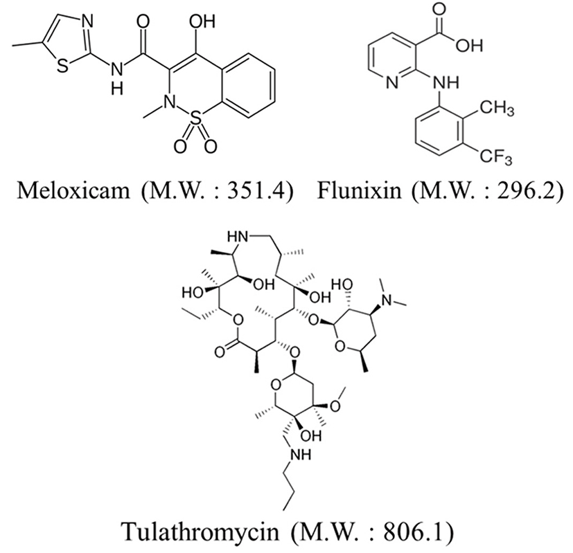
Beef, pork, horsemeat, and milk were collected and tested. The sources of the samples were verified via slaughter certificates and certificates of origin issued by distributors. The samples, collected in proportion to the size of the local population, were obtained from Korea’s main consumer areas, metropolitan cities (Seoul, Busan, Incheon, Daegu, Gwangju, Daejeon, Ulsan and Jeju), and Jeju based on the data provided by the Ministry of Public Administration and Security. A total of 152 samples (47 of beef, 72 of pork, 3 of horsemeat, and 30 of milk) were collected. The edible parts (loins and pasteurized milk) of the collected samples were ground, homogenized, and stored in a 20℃ freezer before analysis. The standards of meloxicam, flunixin, and tulathromycin were purchased from Dr. Ehrenstorfer GmbH (Germany) and their chemical structures are shown in Fig. 1.
Flunixin D3 was used as an internal reference material for both meloxicam and flunixin whereas azythromycin was chosen as the internal standard for tulathromycin. These reference compounds were also purchased from Dr. Ehrenstorfer GmbH (Germany). Meanwhile, methanol and acetonitrile were purchased from Merck Inc. (Germany) for Class High-Performance Liquid Chromatography (HPLC). In addition, other special experimental drugs were also used.
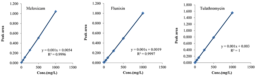
Suitable amounts of meloxicam, flunixin, and tulathromycin (reference materials) and flunixin D3 and azythromycin (internal reference materials) were weighed and dissolved in methanol to give a 1,000 mg/L standard stock solution. For the standard solution of reference materials, the stock solution was diluted with methanol to the desired concentration. The internal standard solutions of flunixin D3 and azythromycin were prepared at 0.05 mg/L. The prepared solutions were stored in brown bottles (in a 4℃ refrigerator) and diluted immediately before use. For each reference material, the calibration curves were generated using concentrations of 0.02, 0.05, 0.10, 0.50, and 1.00 mg/L (Khurana et al., 2013).
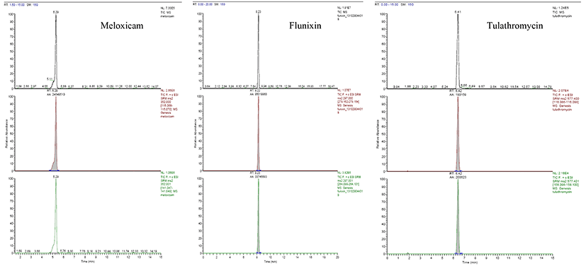
For the Liquid Chromatography-Tandem Mass Spectrometry (LC-MS/MS) experiment, a Thermo Fisher TSQ Quantum Ultra mass spectrometer (Thermo Electron Co., USA) was used, and the system was coupled to a Capcell Pak C8 120 (2.0×50 mm, 5 μm, Imtakt, USA) column. Ammonium formate solution and acetonitrile were used as the mobile phase to analyze meloxicam and flunixin, whereas tulathromycin was analyzed using water and acetonitrile (both containing 0.1% formic acid) using a gradient elution as shown in Table 1. To establish the optimum mass analyzer condition, each reference material was analyzed directly with a tandem mass analyzer without passing through the column. Using the positive ion mode of the Electro-Spray Ionization (ESI) method, the precursor ion for each substance was selected and the collision energy was optimized. The detection condition was set to Multiple Reaction Monitoring (MRM) mode to determine the qualitative and quantitative ions (Yue et al., 2007).
The analyses of meloxicam and flunixin were carried out according to the methods of Korean Food Standards Codex. For dairy products, a homogenized sample (2 g) was introduced into a centrifugation tube, before adding 50 μL and 15 mL of the standard solution and acetonitrile respectively. The mixture was shaken for 10 min and subsequently, Sorvall Legend X1R centrifuge (Thermo Electron Co., USA) at 15,000 rpm for 10 min. The supernatant liquid was taken and the solvent was dried with nitrogen gas below 60℃. Next, the residue was dissolved in a 1 mL mixture of water and methanol (10/90, v/v) and its pH was adjusted to 2.0 using acetic acid. This solution was absorbed onto an HLB cartridge, which was first preactivated with 1 mL of methanol and water each, and then rinsed with 1 mL of 5% methanol solution and finally flushed with another 3 mL of methanol. The solvent of the effluent solution was dried with nitrogen gas below 60℃ and the residue was dissolved in 200 μL of methanol to be used as a test solution. For other types of products, a homogenized sample (5 g) was introduced into a centrifugation tube, before adding 50 μL and 15 mL of internal standard solution and acetonitrile, respectively. The mixture was shaken for 30 min and centrifuged at 15,000 rpm for 10 min. The supernatant liquid was taken and the solvent was dried with nitrogen gas below 60℃. Next, the residue was dissolved in 3 mL of 50% methanol solution. This solution was absorbed onto an HLB cartridge, which was pre-activated with 1 mL of methanol and water each, and then rinsed with 1 mL of 5% methanol solution and finally flushed with another 3 mL of methanol. This effluent was nitrogen-enriched at 60℃ and the residue was dissolved in 200 μL of methanol to be used use as a test solution.
The analysis of tulathromycin was carried out according to the methods of analysis provided in the Korean Food Standards Codex. An internal reference material (azythromycin, 5 mg/L, 1 mL) was added to a 10 g sample, followed by the addition of 10 mL of acetonitrile. The mixture was homogenized for 5 min and then centrifuged at 4℃ and 3,500 rpm for 20 min. The supernatant liquid was collected in a nitrogen fuzzy tube and 10 mL of acetonitrile was added to the sample. The mixture was shaken for 30 s and homogenized for 5 min. It was then centrifuged at 4℃ and 3,500 rpm for 20 min. The supernatant liquid was mixed with the previous batch of supernatant liquid and then dried at <50℃ of nitrogen current. Subsequently, the concentrated residue was dissolved in 3 mL of 2% aqueous formic acid solution, before adding 500 μL of hexane. The solution was first homogenized for 1 min, and then centrifuged at 4℃ and 13,000 rpm for 20 min. The hexane layer was then removed to extract this fluid. The extract was absorbed onto an HLB cartridge, which was pre-activated with 3 mL of methanol and water each, rinsed with 3 mL of 2% aqueous formic acid solution, and then flushed with 3 mL of methanol (containing 2% ammonium hydroxide). The effluent was dried by nitrogen gas at temperatures <50℃ of and the residue were dissolved in 2 mL of distilled water, leaving the solution to homogenize for 1 min. The solution was centrifuged at 4℃ and 13,000 RPM for 20 min and then filtered with a 0.45-μm membrane filter to be used as a test solution.
Prior to actual application, the test methods were verified according to the Korean Food Standards Codex’s method validation guidelines for the detection of veterinary drug residue as provided by Codex Standards. For meloxicam, flunixin, and tulathromycin, their respective calibration curves were generated at concentrations of 0, 0.02, 0.05, 0.10, 0.50, and 1.00 mg/kg. As for the recovery factor, the average and relative standard deviation (RSD) of the samples were calculated from experimental triplicates based on the MRL concentration stated in the Korean Food Standards Codex for each compound. For an analysis of the recovery factor, the drugs were added to each sample at a level of the standards and a test on the recovery factor was conducted according to the methods provided in the Korean Food Standards Codex. The average and relative standard deviation of the samples were calculated via experimental triplicates at each concentration (Wasfi et al., 1998).
Results and Discussion
The conditions of LC-MS/MS analysis to establish the limits of detection and quantitation were set according to the veterinary drug residue test methods specified in the Korean Food Standards Codex. The instrumental conditions used to analyze each substance are shown in Table 4 and their respective calibration curves were generated using different standard concentrations. The limit of quantitation was calculated with a signal-to-noise (S/N) ratio of over 10 and as a result, the limit of quantitation was 0.0003-0.0005 mg/kg.
| Beef | Pork | Milk | Horse-meat | Total | |
|---|---|---|---|---|---|
| Seoul | 15 | 18 | 10 | 43 | |
| Busan | 10 | 16 | 7 | 33 | |
| Inchon | 6 | 9 | 4 | 19 | |
| Daegu | 5 | 8 | 3 | 16 | |
| Gwangju | 4 | 7 | 2 | 13 | |
| Daejeon | 4 | 7 | 2 | 13 | |
| Ulsan | 3 | 7 | 2 | 12 | |
| Jeju | 3 | 3 | |||
| Total | 47 | 72 | 30 | 3 | 152 |
a)ND, Not Detected (below the detection limit); b)D, Detected.
For a test on the recovery factor of meloxicam, flunixin, and tulathromycin, appropriate reference materials were added to the beef, pork, and milk samples (i.e., 0.02 mg/kg for beef, 0.02 mg/kg for pork, and 0.015 mg/kg for milk) according to the test methods specified in the Korean Food Standards Codex. The recovery factor of meloxicam was 89.8-92.9% whereas the RSD was 1.5-7.7%, both of which meet the standards required by the Codex (i.e., recovery factor at 70-120% and RSD <20%) (Table 2). Meanwhile, the recovery factor of flunixin was 89.8-92.9% and the RSD was 1.5-7.7%, both of which also meet the standards required by the Codex (i.e., recovery factor at 70-120% and RSD <20%). Lastly, the recovery factor of tulathromycin was 87.2-89.8% and the RSD was 2.6-6.1%, and both sets of values meet the standards required by the Codex (i.e., recovery factor at 70-120% and RSD <20%).
In 1989, 40 MRLs of veterinary drugs were established by the Ministry of Food and Drug Safety and this original list has been expanded to include 156 MRL values to date; with a history of active enforcement since 2006 (Ministry of Food and Drug Safety, 2013). Since veterinary drugs are medicines used on living organisms, the frequencies of their use or occurrence can only be analyzed with practical methods. As such, a survey on the actual conditions of veterinary drug residues contains more risks than anything else. Thus, this survey on the actual situation of veterinary drug residues found in animal products was performed to accumulate scientific data, which could be used for forecasting and safety management. Food samples were collected from seven major metropolitan cities nationwide, along with Jeju, and their sources were thoroughly verified through slaughter certificates and certificates of origin issued by the distributors. Tulathromycin was found in five samples (4.2%) out of 119 samples of beef and pork found to contain veterinary drug residues and 5 samples were from pork sources. When analyzed according to the sampling area, two samples were found in Seoul and three in Daegu (Table 5). All residues were detected below the stipulated MRLs and tulathromycin was detected at 0.1 mg/kg in the five aforementioned samples.
| Region | Sample (Pork) | Level (mg/kg) | MRLs (mg/kg) | |
|---|---|---|---|---|
| Tulathromycin | Seoul | P-11 | 0.0013 | 0.1 |
| Seoul | P-12 | 0.0012 | ||
| Daegu | P-137 | 0.0011 | ||
| Daegu | P-138 | 0.0010 | ||
| Daegu | P-139 | 0.0013 |
This study analyzed the presence of drug residues in beef, pork, horse-meat, and milk sources that are circulated domestically by post-monitoring three different kinds of veterinary drugs, including meloxicam, flunixin, and tulathromycin. The results are used as a basic guideline to develop food safety policy, to verify the test methods provided in the Korean Food Standards Codex, and to check residual level for legal compliance. Tulathromycin was detected in five samples and two samples were from Seoul with another three from Daegu. These were detected in pork sources meant for domestic consumption and the samples were all found to have MRL values below 0.1 mg/kg. A test of recovery factors was conducted according to the test methods of veterinary drug residue specified in the Korean Food Standards Codex (n=3). According to the test results, recovery factors of 82-98% and RSD of 1.5-7.7% were obtained and these values meet the standards required by the Codex (recovery factor of 70-120% and RSD <20%). In this study, five veterinary drugs were detected in a total of 152 samples, giving a detection rate of approximately 3.3%; with no violation of legally acceptable values. While the animal products analyzed are considered safe for consumption, a more persistent and widespread monitoring, along with institutional management of veterinary drugs would be necessary to ensure consumer food safety and effective management of veterinary drugs.
Conclusions
For this survey of 3 kinds of veterinary drugs, monitoring of 152 samples were analyzed by liquid chromatography- tandem mass spectrometry according to the Korean Food Standards Code. Tulathromycin was found in five samples of pork sources. All residues were detected below the stipulated MRLs was detected at 0.1 mg/kg. Even though detection level was below the MRLs established for food safety management, still continuous and extensive monitorning is required in the near future.