


Introduction
Sodium chloride is the main ingredient for processing meat products. It provides not only desirable textural properties and flavor, but also enhances water and fat binding ability during cooking (Ruusunen and Puolanne, 2005). However, consumption of large amounts of dietary sodium chloride causes high blood pressure, thus increasing the risk of cardiovascular disease (Tuomilehto et al., 2001). For this reason, studies on low-salt meat products have been conducted in order to reduce the risk of these diseases. The purpose of these researches is to reduce contents of salt and prevent detrimental effects such as low textural properties, water-holding capacity, and sensorial attributes.
Gelatin is a by-product produced by partial hydroloysis of collagen extracted from animal and fish. Its hydrocolloid can be used as gelling and water-binding agent. Peptide chains of gelatin are composed of glycine, proline, and hydroxyproline that can increase the water-holding capacity. Especially, the content of hydroxyproline in gelatin is highly associated with gel strength (Sarabia et al., 2000). Pork skin gelatin used in the present study has higher amounts of glycine, serine, tyrosine, and proline than bovine skin gelatin. Gel strength of pork skin gelatin is also superior to that of bovine skin gelatin (Nur Hanani et al., 2014). Water holding capacity of pork skin gelatin at pH 6 is also higher than that of fish or bovine gelatin (Koli et al., 2013).
This study might figure out that gelatin can reduce the detrimental effects, which caused by reducing the salt concentrations, in meat products. To investigate the effect of gelatin in meat at low salt concentrations, the objective of this study was to evaluate the effect of different salt concentrations on rheological properties of myofibrillar protein (MP) gel containing pork skin gelatin.
Materials and Methods
Fresh pork loin (Crossbred pig by Landrace×Yorkshire, Grade A) was bought from a local market. After trimming all visible fat and connective tissues, it was cut into cubes (2 cm3) and stored at −50°C freezer until analyzis. Pork skin gelatin powder was obtained from Gel-Tech (Gelatin-G, Busan, Korea). This gelatin powder had 209 bloom of jelly strength and 8 mesh of particle size.
Pork loin was ground to emsulsions with 50 mM phosphate and 0.1 M sodium chloride solution. After centrifuging at 1,590×g for 15 min, the precipitate was homogenized with 0.1 M sodium chloride buffer solution and connective tissue in the emulsion was removed. MP was adjusted to 4% (protein concentration) by adding buffer solution. MP mixtures were then prepared with or without 1.0% of gelatin powder at different salt concentrations (0.15, 0.30, and 0.45 M) as shown in Table 1. MP mixtures were then loaded into vial tubes and heated from 20°C to 80°C at 3°C/min increments. Cooked samples were cooling down in an ice water bath and then stored in a refrigerator until utilized.
Cooking yield (CY, %) was calculated based on weight difference beween weight before cooking and weight after cooking. Results were obtained by averaging five measurements of each sample. After cooking, gel strength of sample was measured using an Instron Universal Testing Machine (Model#3344, Canton, MA, USA). The probe head speed was set at 500 mm/min and breaking force (gf) was recorded.
MP mixtures were loaded into the container of a probe on a rheometer. Shear stress was measured using a concentric cylinder type rotational rheometer (Model #RC30, Rheotec Messtechnik GmbH, Ottendorf-Okrilla, Germany). The shear rate ranged from 0 to 600/s.
Intensities of protein bands from MP containing gelatin and different salt levels were determined. Acrylamide gel was made with 10% separating gel and 4% stacking gel (Laemmli, 1970). Samples were diluted to have 1% protein concentration. Diluted samples were then mixed with sample buffer before loading to the separating gel. After proteins were separated at 150 V for 1.5 h, the acrylamide gel was stained with Coomassie brilliant blue staining solution and then destained. The gel was then analyzed compared to a standard protein marker (Model#161-0318, Bio-Rad, CA, USA).
Samples with a small cube shape (3 mm3) were soaked overnight in 2.5% glutaraldehyde solution in a refrigerator (4±1°C). After samples were incubated with osmium tetroxide solution at room temperature for 5 h, they were dehydrated with increasing concentrations of ethanol (50%, 60%, 70%, 80%, 90%, and 100%) and acetone. Samples were then gold-coated using a coating machine (Model#108 autos putter coater, Cressington Scientific Instruments Ltd., Watford, UK). Coated samples were observed with a LV-SEM (Model#JSM-6610LV microscope, JEOL Ltd., Tokyo, Japan).
FTIR samples were prepared the same as aforementioned in LV-SEM. Their secondary structures were determined with a Fourier transform infrared spectroscope (Frontier FT-IR/NIR Spectrometer, PerkinElmer, Waltham, MA, USA). Scanning wavelength ranged from 4,000 to 400 cm−1.
DTNP [2,2’-dithiobis(5-nitropyridine)] solution was used to determine sulfhydryl contents with modified Ellman’s method (Sun and Holly, 2011). Briefly, 0.1 g MP sample was incubated in a solution mix of 0.5 mL DTNP solution, 1 mL Tris, and 8.4 mL distilled water. Its absorbance at 412 nm was then measured with a spectrophotometer (UV-1601, Shimadzu, Kyoto, Japan).
Protein surface hydrophobicity was analyzed using hydrophobic chromophore bromophenol blue (BPB) solution (Chelh et al., 2006). Briefly, 1 g of each sample and BPB 0.5 mL were vortexed for 10 min. A 20 mM phosphate buffer solution was used as a control. The mixture was centrifuged at 1,660×g for 15 min and absorbance of each supernatant was measured at 595 nm using a spectrophotometer (UV-1601, Shimadzu). Values of protein surface hydrophobicity were calculated using the following formula:
Two-way (2 levels of gelatin×3 levels of salt conc.) analysis of variance (ANOVA) was used to analyze CY and gel strength. To analyze sulfhydryl contents and protein surface hydrophobicity, one-way ANOVA was performed. Each experiment was performed in triplicate. Significant difference was considered at p<0.05.
Resutls and Discussion
Table 2 shows the effect of different salt concentrations on CY and gel strength values of MP gels containing 1% gelatin. The addition of gelatin powder increased the CY (p<0.05). The addition of gelatin also increased the length of polypeptide chain with hydroxyl groups, resulting in increased CY of MP gels (Sun and Holley, 2011). These polypeptide chains in triple helix were repeated with glycine, proline, and hydroproline, indicating that these amino acids had hydrophobic properties, thus entrapping free water among meat structures (Nur Hanani et al., 2014). MP gel at salt concentration of 0.15 M had lower CY than that at higher salt level of 0.30 M or 0.45 M. The addition of sodium chloride caused repulsion induced by negative charges, resulting in swelling of myofibrils and enhanced water-holding capacity (Desmond, 2006). The addition of salt increased concentrations of electrolyte that could reduce the thickness of diffusion double membrane of gelatin surface. Thus, gelatin is easily aggregated to form large aggregate which affects water holding capacity (Sow and Yang, 2015).
The gel strength of MP gel at the salt concentration of 0.45 M was higher than that at salt concentration of either 0.15 or 0.30 M regardless of gelatin addition. However, no significant difference in gel strength (p>0.05) between 0.15 and 0.30 M salt concentrations was observed. The addition fo gelatin did not significantly increase the gel strength partially due to the the lack of cystein and cystin in gelatin which could not form disulfide bond for protein gelation, resulting in no effect on gel strength of MP gel (Sun and Holley, 2011). The lower length chain of gelatin might form weak gel because of lower junction zones among structures (Benjakul et al., 2009). Gel formation of MP with gelatin did not significantly improve the gel strength since they were not combined well with each other. They formed MP and gelatin gelation, respectively. The addition of sodium chloride might have broken down hydrogen bonds of gelatin and interferes with hydrophobic interaction (Choi and Regenstein, 2000). Storage modulus of gelatin decreased with increasing level of salt because of the screening off of short-range electrostatic interaction (Haug et al., 2004). Salt-soluble MP extracted with the addition of salt on meat protein could strongly bind among proteins, resulting in increased gel strength of MP gels (Desmond, 2006).
Shear stress values of MP mixtures with gelatin at different salt levels are shown in Fig. 1. The shear stress had quite difference among treatments with and without gelatin. The shear stress of MP mixtures without gelatin at 10 (1/s) shear rate ranged from 8 to 84 (Pa). On the contrary, shear stress of MP mixtures with gelatin ranged from 166 to 478 (Pa) at below 10 (1/s) shear rate. It might be affected by the high molecular weight of peptide chain from pork skin gelatin, indicating that high viscosity could induce tough and extensible gelling properties (Rafieian et al., 2015). The amount of serine which has free hydroxyl groups in pork skin gelatin can bind with water molecular by hydrogen bonds, resulting in gelatinous state of gelatin in MP mixtures that can increase shear stress (Raja Nhari et al., 2011). The screening of electrostatic interaction could expand gelatin by the addition of salt (Qiao et al., 2013). Yang et al. (2007) have reported that elastic modulus of gelatin is higher than that of myosin, indicating that electrostatic from interaction of myosin and gelatin can induce highly elastic gel. MP mixtures at salt concentration of 0.45 M had higher shear stress values than those at other lower salt concentrations (0.15 and 0.30 M). Bertram et al. (2004) have reported that MP with increasing ionic strength has high water-binding capacity, thus inducing swelled structure by electrostatic repulsion.
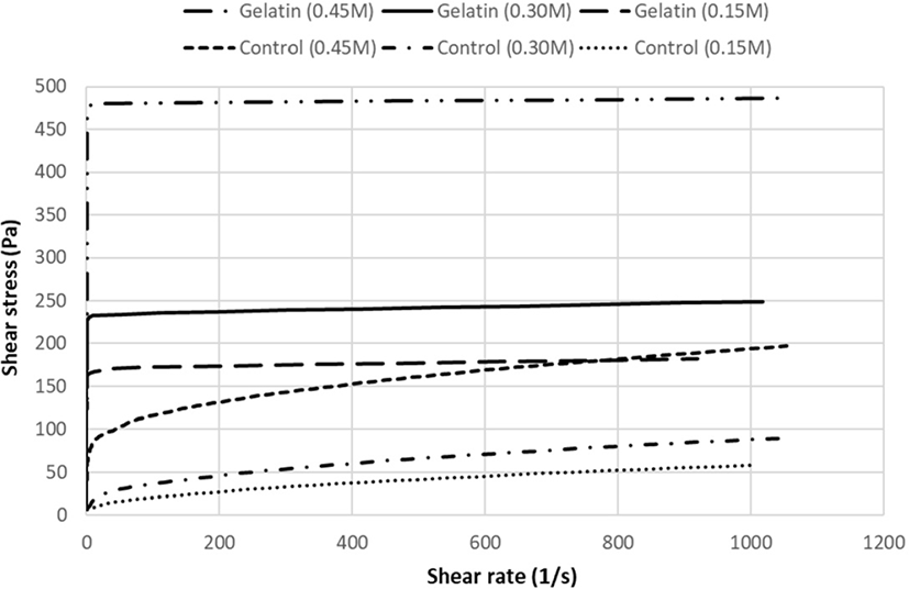
Fig. 2 shows SDS-PAGE of MP mixtures with gelatin at different salt levels. Protein pattern added with gelatin showed bands at above 195 kDa. In accordance with this result, Raja Nhari et al. (2011) have shown a protein band of gelatin at high molecular weight. Intensities of myosin heavy chain (MHC) and actin were also decreased with the addition of gelatin and increasing levels of salt. Reduction of protein intensity has been observed by protein cross-linking as hydrogen bond and hydrophobic interactions (Nuanmano et al., 2015). Therefore, MP mixtures incorporated with gelatin and increasing levels of salt produced high molecular weight of protein that could form aggregated protein matrix, thus inducing gelation and water-holding capacity.
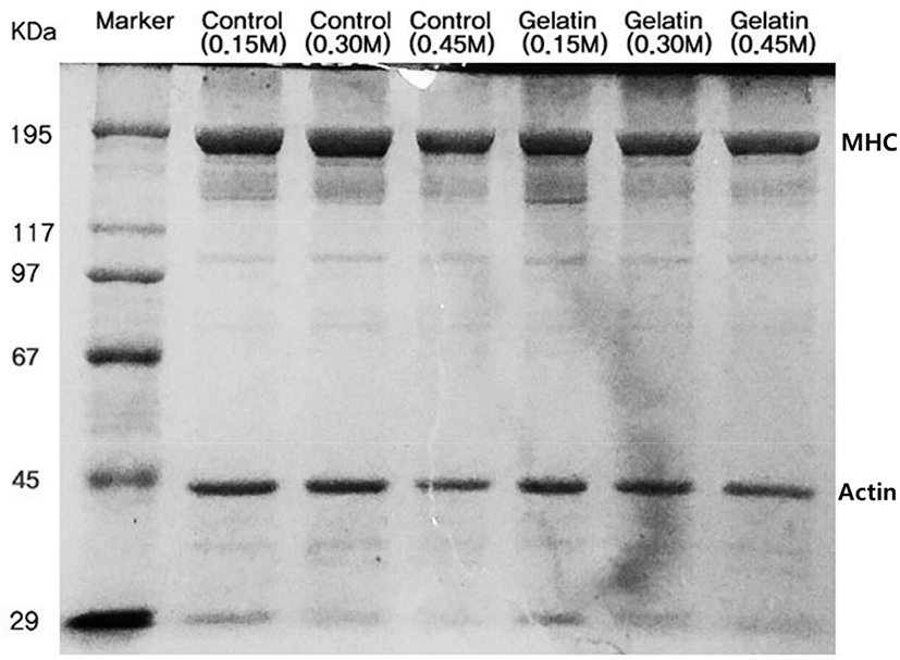
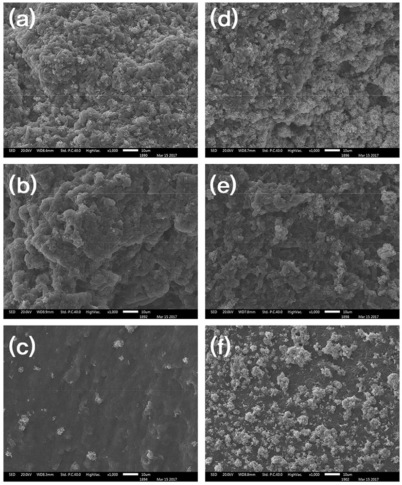
Fig. 3 shows microstructures of MP gels with gelatin at different salt levels. MP gels with increasing salt concentration showed more uniform structures with small pores. At low salt concentration, MP gels showed irregular microstructures. Among structures at different salt concentrations, porosity related to water-binding capacity showed main difference (Zhang et al., 2015). Sun and Holley (2011) reported that the microstructure of MP gels at low-ionic strength showed fine-stranded gel structure, and MP gels at high-ionic strength showed the coarsely aggregated structures. When MP gels at 0.45 M salt concentration with and without gelatin were compared, the addition of gelatin showed gelatin particles evenly attached to a uniformed structure.
Quantities of α-helix and β-sheet in gelatin were higher than those in MP gels containing gelatin at different salt concentrations as shown in Table 3. Quantitative analysis of changes in paeks at 1,650 cm−1, 1,624 cm−1, and 1,680 cm−1 showed that the quantity of α-helix/unordered structures and β-sheet was decreased with increasing salt concentrations. According to study by Bertram et al. (2004), salt-soluble proteins were folded during heating of MP. Thus, MP gels with increasing sodium chloride concentration showed decreased quantity of secondary structures. When gelatin was added to MP gels, no significant difference in gel strength was observed. The quantity of secondary structures was the highest for gelatin powder which was composed of α-chains, β-chains, and γ-chains (Nur Hanani et al., 2014). The addition of sodium chloride to gelatin blocked the short-range of electrostatic interactions, resulting in reduction of bridge on α-chains (Haug et al., 2004). The secondary structure of gelatin was decreased with addition of sodium chloride (Sow and Yang, 2015). There is a negative correlation between MP gel strength and α-helix content. Unfolded α-helix and β-sheet structure might have been converted to a tertiary structure to increase the gel strength of MP.
Sulfhydryl contents of MP mixtures with gelatin at different salt levels are shown in Table 4. Increasing salt concentration lowered the content of sulfhydryl groups but enhanced the solubility of MP, resulting in exposing buried sulfhydryl groups on internal structures and formation of disulfide bonds (Zhang et al., 2015). On the contrary, MP mixture without gelatin or with gelatin had no difference in sulfhydryl content at each salt level (p>0.05) since gelatin had no cysteine or cystine in their compositions for disulfide bonding (Sun and Holley, 2011). Although gel formation of gelatin was not influenced by disulfide bond, junction zones of gel network were bound by electrostatic interaction and hydrogen bonds (Haug et al., 2004).
Table 4 shows protein surface hydrophobicity of MP mixtures with gelatin by different salt levels. Protein surface hydrophobicity of MP at 0.45 M was higher than that at lower salt concentration (<0.3 M) (p<0.05). As protein interactions with increasing salt concentrations were weakened, hydrophobic groups were exposed on the surface of MP (Zhang et al., 2015). At each level of salt concentration, MP mixture with gelatin had higher hydrophobicity value than that without gelatin (p<0.05). As gelatin is consisted of many amino acids with hydrophobicity, MP mixtures incorporated with gelatin also showed high protein surface hydrophobicity values (Nur Hanani et al., 2014).
Conclusion
The addition of pork skin gelatin into MP gels increased the CY and induced high protein surface hydrophobicity and polymerization. Salt concentrations at 0.30 M and 0.45 M improved the CY of MP gels compared to that at 0.15 M. However, gel strength was not affected by adding gelatin. Salt cocentration at 0.45 M had superior effect due to more sulfide bonds and higher hydrophobicity. Even at low salt concentration, the addition of gelatin increased CY and viscosity of MP gels and it might improve physicochemical properties in low-salt meat products.