


Introduction
Foot-and-mouth disease (FMD) is a highly contagious disease that affects both wild and domestic cloven-hoofed animals, including cattle, sheep, goats, and pigs, and can lead to economically devastating outcomes (Parida, 2009). FMD limits access to markets for developing countries and has huge costs related to testing, slaughter, vaccination, production losses, transport restrictions, and so on (OIE, 2012), as evidenced by the costs of outbreaks in the UK and the Netherlands in 2001 and again in the UK in 2007. The causative agent of FMD is the FMD virus, which is a single-stranded positive-sense RNA virus belonging to the genus Aphthovirus in the family Picornaviridae (Parida, 2009).
A 2011 outbreak of FMD in South Korea rapidly affected the whole country, and the government adopted a nationwide vaccination program in response to this emergency. Although the vaccine was administered at 8 wk of age, the antibody titers against the FMD type O virus were not sufficiently high and there were side effects, or “adverse reactions”, including abscesses and/or granulomas formed at the inoculation sites. Although most vaccines cause some local side effects, such as pain, redness, swelling, or a small lump at the injection site, these usually resolve shortly (Gerlach et al., 2012; Van den Broeke et al., 2016). However, there is a well-documented history in meat inspection reports of local reactions at the inoculation site in pigs vaccinated with FMD antigens formulated with oil adjuvant (Batista-Duharte et al., 2013). Oil and aluminum hydroxide adjuvant have long been used in human and veterinary intramuscular inoculations. However, the abscess or granuloma that develops where the injection is given often does not disappear by slaughter. As a result, a portion of the muscle has to be removed, creating an economic loss (Vatulini et al., 2005; Gerlach et al., 2012). Because adipose tissue or fascia lack of blood supplies, absorption is delayed and abnormal meat can be formed when the vaccine is not injected into the muscles precisely. In addition, the irritating components of the vaccine as well as contaminated needles can contribute to tissue necrosis and inflammation at the injection site.
For these reasons, this study aimed to investigate the economic impact of abscess or granuloma formation at the foot-and-mouse disease vaccine (FMDV) injection site on the swine industry and to evaluate the effect of injection device, i.e., syringe-needle vs needle-free jet injector, on the incidence of abscesses and granulomas. Additionally, morphometric and histopathological examinations were conducted on the abnormal meats. Because the Korean government adopted a nationwide FMD vaccination program after the FMD outbreak in 2011, the clinical signs of granuloma formation at the injection site in experimental pigs as well as FMD vaccinated slaughtered pigs from a commercial farm were simultaneously monitored at a slaughter house in 2010–2015.
Materials and Methods
A total of 276 colostrum-fed, cross-bred, conventional piglets were selected and purchased from a 9 different commercial farms in Dodram Pig Farmers’ Cooperative at 35 d of age. Sows on the farm were vaccinated against FMD at 4–5 wk before farrowing. All of the pigs were housed within pens in an environmentally controlled building with completely slatted floors throughout the experimental period. The pigs underwent a 21-day conditioning period at the beginning of the study.
The animals were randomly divided into three groups as follows: a conventional “syringe-needle vaccination into the neck” group (N-Neck, n=71), a “syringe-needle vaccination into the ham” group (N-Ham, n=70), and a “needle-free jet injection into the neck” group (Non-Neck, n=135). At 56 d of age, the piglets were vaccinated with 2 mL of a commercially available FMD vaccine (Decivac®, Intervet, Germany), which is a trivalent vaccine containing serotypes O, A, and Asia 1 emulsified with the adjuvant Montanide ISA206 (W/O/W, Seppic, France). The vaccine was inoculated intramuscularly (IM) on the left side of each animal (left neck or ham) and lesions if any at left side of injection site were monitored. All treatments, except for the FMD vaccine were injected IM on the right side to differentiate between FMDV from other vaccinations. The inoculation area was thoroughly cleaned with Betadine® (a complex solution of iodine and ammonium quaternary salts), ethyl alcohol, and Betadine® to avoid any iatrogenic bacterial infection (Vatulini, 2005). All groups were maintained without other treatments, except minimum vaccinations. At slaughter, government inspectors identified carcasses with abscesses/ granulomas at the injection sites, and the condemned parts were examined according to the standardized inspection process at the slaughter facility. The pigs used in this study were cared for according to the guidelines of the Institutional Animal Care and Use Committee of Kangwon National University.
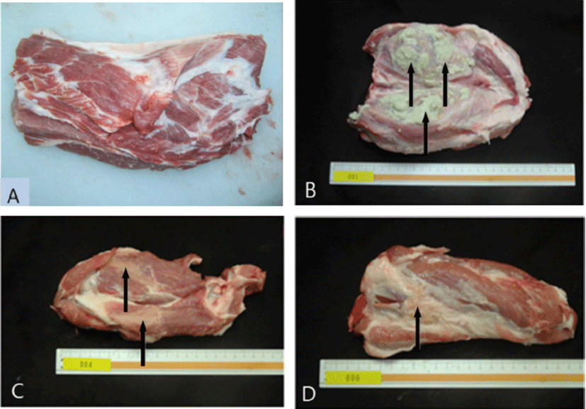
Morphometric analyses of the gross and microscopic lesions at injection sites were conducted (Fig. 1) at the slaughter facility to see if there was a “severe abscess” or “discoloration of the muscle and granuloma” as previously described (Vatulini, 2005). In brief, any lesion found at slaughter was collected and sent to a laboratory for microscopic examination. Granulomatous lesions appeared as protruding, firm nodules located within the muscle tissue and subcutaneous fat. When cut, a hardly encapsulated white-yellowy material was observed. Sometimes, a series of small nodular-capsulated formations were also observed scattered throughout the muscle tissue.
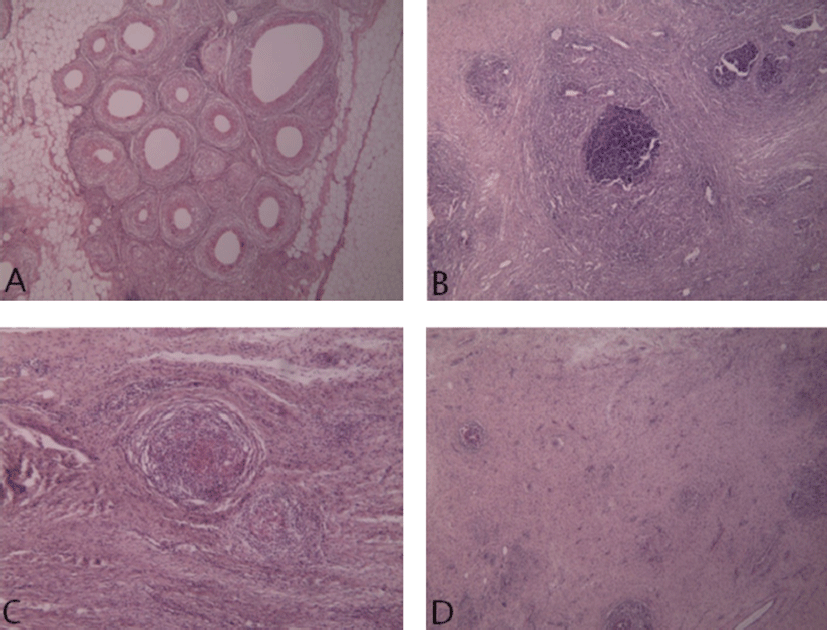
For the histopathological evaluation, tissue samples were collected, fixed in 10% (w/v) neutral-buffered formalin for 24–48 h, and embedded in paraffin wax. Sections (4 μm) were stained with hematoxylin-eosin for light microscopy (Fig. 2). The lesions were examined and sorted according to the following four criteria: “vaccine remaining”, “abscess”, “granuloma” and “fibrosis.” Histological pictures were taken of all lesions, and the focal granulomatous tissue was evaluated for chronic inflammation (abscess), aggregates of macrophage-derived epithelioid cells with oval nuclei (vaccine remaining), and pale pink cytoplasm and indistinct cell borders (granuloma and fibrosis) as described by Parker et al. (2009).
The injection site in each pig was visually examined, and the incidence of a granuloma was confirmed. After trimming the affected area, it was weighed and multiplied by the weekly sales price obtained from Korea Pork Producers Association to calculate the economic loss.
Data were analyzed using the generalized linear model procedure (PROC-GLM) of SAS (SAS Institute, USA, 2009). Differences among treatment groups were determined by using Duncan’s new multiple range tests. Discrete data for the occurrence of gross and histopathological lesions were calculated as the percentage of affected carcasses in each group and were analyzed by Chi-square and/or Fisher’s exact test. A p-value less than 0.05 was considered statistically significant.
Results and Discussion
Severe abscesses at the injection sites were most frequently observed in the Non-Neck group, followed by the N-Neck and N-Ham groups (p<0.05, Table 1). “Discoloration of muscle and granuloma” was the highest in the N-Neck group, followed by the Non-Ncek and N-Ham groups (p<0.05, Table 1, Fig. 1).
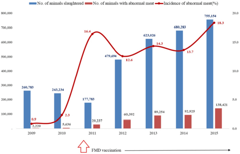
Because the Korean government adopted a nationwide FMD vaccination program in 2011, we continuously monitored not only the pigs those in our experimental groups but also FMD vaccinated pigs from Dodram Pig Farmers’ Cooperative at a slaughter house in 2009–2015 (n=3,219,957, Fig. 3). In the period before implementation of the vaccination program (2009–2010), the average granuloma/abscess occurrence rate was 1.6%. However, after implementation of the FMD vaccination program, the rate increased sharply (p<0.05) to 15.1% (Fig. 3), suggesting granulomas and abscesses as a side effect of FMD vaccination on pork quality.
The incidence rates of “vaccine remaining”, “abscess”, “granuloma” and “fibrosis” were significantly lower (p<0.05) in the N-Ham group than in the other two groups (N-Neck and Non-Neck, Table 2).
There were significant differences (p<0.05) among the groups in total condemned meat weight, condemned meat per pig, total economic loss, and economic loss per pig. Overall, the Non-Neck group had higher economic losses than the other two groups (p<0.05, Table 1).
The occurrence of FMD type O in Korea in 2010 triggered a nationwide vaccination program. However, the potency of the vaccine against FMD as well as the adverse reactions induced by vaccination and their effects on carcass quality warrant further evaluation.
Muscular lesions at the sites where drugs and vaccines are inoculated can lead to potential economic losses in the livestock industries (Vatulini, 2005), and reports of local reactions after inoculation of the FMDV emulsified in double oil adjuvant in pigs have long been a concern (McKercher, 1971). The serologic data indicate that either the subcutaneous or intramuscular route, as well as the different inoculation sites selected showed a similar FMDV efficacy in terms of antibody response (McKercher, 1971). Because there is no doubt about vaccine efficacy on different inoculation sites and sales price difference on pork cuts, we compared the economic losses caused by FMDV on different injection sites.
Before FMD vaccination, the economic losses related to local inoculation reactions in Korea was approximately US $28 per pig (Kim et al., 2012), for a total of $255,000 in 2010. However, post-FMD vaccination (in 2011), these losses increased remarkably to a total $1,673,000. Based on our field study of pigs from Dodram Pig Farmers’ Cooperative, the average yearly total economic losses due to abnormal meat in the neck area before and after the FMD vaccination program were $309,124 and $1,056,088 in 2009 and 2015, respectively. Therefore, it is obvious that the injection route for the FMDV should be reconsidered to minimize the incidence of abnormal pork. At the slaughter house, vaccine remnants were observed as encapsulated granulomas containing material engulfed by macrophages, epithelioid cells, giant cells, and granulocytes or eosinophils, which are the cardinal signs of chronic inflammation, as described by Chong et al. (2006). The neck area of a pig contains several brachiocephalic muscles. If the injection is administered close to the shoulder, the material could easily permeate in between the myofascial areas, which lack absorbing capability. Then, the injected material could be remain for a long period of time, leading to chronic activation of antigen presenting cells, such as macrophages and dendritic cells, resulting in lymphocyte infiltration (abscess). Our data showed that the N-Ham group presented significantly fewer injection reactions than the N-Neck and Non-Neck groups. We speculate that the ham area contains relatively larger and wider muscle components than the neck area, making the injection more precise. Additionally, the results suggest that the incidence of granulomas/abscesses in the conventional syringe-needle and needle-free injector groups was not different, which is similar to previous studies of injection devices (Houser et al., 2004; King et al., 2010). In fact, when we designed the experimental groups for this study, we had planned to add a fourth group in which the vaccine was inoculated into the ham area using a needle-free injector. However, because more labor is needed to hold the animal still for precise positioning of the injector into the ham, which is not easy, especially at this age, this experimental group, needle-free jet injection into the ham route, was not included.
In summary, the economic losses from injection site granuloma/abscess formation were significantly higher post FMD vaccination. The economic losses caused by removing the condemned muscular portions from the area with granulomas were identical in all affected pigs, but the number of lesions could be reduced if the ham route was employed for vaccination. Vaccines approved for use in animals as well as the inoculated areas in animals consumed as food by humans need to be evaluated carefully.